Translate this page into:
A rare case of spotted grouped pigmented naevus: Congenital follicular melanocytic naevus
Corresponding author: Dr. Vishvender Singh, Department of Dermatology & STD, Vardhman Mahavir Medical College and Safdarjung Hospital, New Delhi, India. vsyen22@gmail.com
-
Received: ,
Accepted: ,
How to cite this article: Singh V, Srivastava P, Sharma S, Khunger N. A rare case of spotted grouped pigmented naevus: Congenital follicular melanocytic naevus. Indian J Dermatol Venereol Leprol. 2025;91:128-9. doi: 10.25259/IJDVL_1110_2023
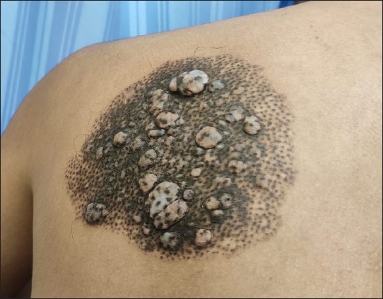
A 24-year-old man presented with a slow-growing, asymptomatic, 20-cm sized well-circumscribed lesion on his left upper back, consisting of several small, follicle-centred, discrete to coalescing, brownish-black to bluish macules. The lesion also demonstrated multiple, soft, well-circumscribed papules and nodules, with overlying follicular pigmentation. There was no hypertrichosis or underlying musculoskeletal defect [Figure 1].

- A well-circumscribed lesion on the back consisting of multiple brownish-black to bluish hyperpigmented macules and multiple soft well-circumscribed papules and nodules.
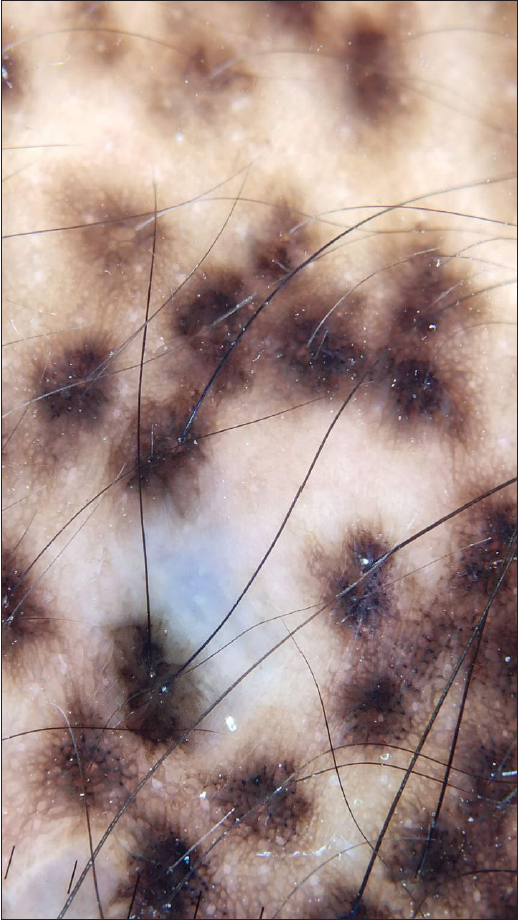
Dermoscopy revealed follicle-centred brownish-black to bluish pigmentation in a reticulo-globular pattern, with intervening normal skin [Figure 2]. On histopathology, the dermis showed aggregates of uniform naevus cells surrounding the hair follicles along with abundant melanophages. Based on clinical features, dermoscopy, and histopathology, we diagnosed it as congenital follicular melanocytic naevus, also called spotted grouped pigmented naevus.

- Dermoscopy showing follicle-centred brownish-black to bluish pigmentation with a reticulo-globular pattern and intervening normal skin (DermLite DL3N; contact, polarised, 10x).
Declaration of patient consent
The authors certify that they have obtained all appropriate patient consent.
Financial support and sponsorship
Nil.
Conflicts of interest
There are no conflicts of interest.
Use of artificial intelligence (AI)-assisted technology for manuscript preparation
The authors confirm that there was no use of artificial intelligence (AI)-assisted technology for assisting in the writing or editing of the manuscript and no images were manipulated using AI.


