Generic selectors
Exact matches only
Search in title
Search in content
Post Type Selectors
Search in posts
Search in pages
Filter by Categories
15th National Conference of the IAOMFP, Chennai, 2006
Abstract
Abstracts from current literature
Acne in India: Guidelines for management - IAA Consensus Document
Addendum
Announcement
Art & Psychiatry
Article
Articles
Association Activities
Association Notes
Award Article
Book Review
Brief Report
Case Analysis
Case Letter
Case Letters
Case Notes
Case Report
Case Reports
Clinical and Laboratory Investigations
Clinical Article
Clinical Studies
Clinical Study
Commentary
Conference Oration
Conference Summary
Continuing Medical Education
Correspondence
Corrigendum
Cosmetic Dermatology
Cosmetology
Current Best Evidence
Current Issue
Current View
Derma Quest
Dermato Surgery
Dermatopathology
Dermatosurgery Specials
Dispensing Pearl
Do you know?
Drug Dialogues
e-IJDVL
Editor Speaks
Editorial
Editorial Remarks
Editorial Report
Editorial Report - 2007
Editorial report for 2004-2005
Errata
Erratum
Focus
Fourth All India Conference Programme
From Our Book Shelf
From the Desk of Chief Editor
General
Get Set for Net
Get set for the net
Guest Article
Guest Editorial
History
How I Manage?
IADVL Announcement
IADVL Announcements
IJDVL Awards
IJDVL AWARDS 2015
IJDVL Awards 2018
IJDVL Awards 2019
IJDVL Awards 2020
IJDVL International Awards 2018
Images in Clinical Practice
Images in Dermatology
In Memorium
Inaugural Address
Index
Knowledge From World Contemporaries
Leprosy Section
Letter in Response to Previous Publication
Letter to Editor
Letter to the Editor
Letter to the Editor - Case Letter
Letter to the Editor - Letter in Response to Published Article
LETTER TO THE EDITOR - LETTERS IN RESPONSE TO PUBLISHED ARTICLES
Letter to the Editor - Observation Letter
Letter to the Editor - Study Letter
Letter to the Editor - Therapy Letter
Letter to the Editor: Articles in Response to Previously Published Articles
Letters in Response to Previous Publication
Letters to the Editor
Letters to the Editor - Letter in Response to Previously Published Articles
Letters to the Editor: Case Letters
Letters to the Editor: Letters in Response to Previously Published Articles
Media and news
Medicolegal Window
Messages
Miscellaneous Letter
Musings
Net Case
Net case report
Net Image
Net Images
Net Letter
Net Quiz
Net Study
New Preparations
News
News & Views
Obituary
Observation Letter
Observation Letters
Oration
Original Article
ORIGINAL CONTRIBUTION
Original Contributions
Pattern of Skin Diseases
Pearls
Pediatric Dermatology
Pediatric Rounds
Perspective
Presedential Address
Presidential Address
Presidents Remarks
Quiz
Recommendations
Regret
Report
Report of chief editor
Report of Hon : Treasurer IADVL
Report of Hon. General Secretary IADVL
Research Methdology
Research Methodology
Resident page
Resident's Page
Resident’s Page
Residents' Corner
Residents' Corner
Residents' Page
Retraction
Review
Review Article
Review Articles
Reviewers 2022
Revision Corner
Self Assessment Programme
SEMINAR
Seminar: Chronic Arsenicosis in India
Seminar: HIV Infection
Short Communication
Short Communications
Short Report
Snippets
Special Article
Specialty Interface
Studies
Study Letter
Study Letters
Supplement-Photoprotection
Supplement-Psoriasis
Symposium - Contact Dermatitis
Symposium - Lasers
Symposium - Pediatric Dermatoses
Symposium - Psoriasis
Symposium - Vesicobullous Disorders
SYMPOSIUM - VITILIGO
Symposium Aesthetic Surgery
Symposium Dermatopathology
Symposium-Hair Disorders
Symposium-Nails Part I
Symposium-Nails-Part II
Systematic Review and Meta-Analysis
Systematic Reviews and Meta-analyses
Systematic Reviews and Meta-analysis
Tables
Technology
Therapeutic Guideline-IADVL
Therapeutic Guidelines
Therapeutic Guidelines - IADVL
Therapeutics
Therapy
Therapy Letter
Therapy Letters
View Point
Viewpoint
What’s new in Dermatology
View/Download PDF

Translate this page into:

Quiz
88 (
6
); 794-796
doi:
10.25259/IJDVL_493_20
pmid:
33969662

A skin-colored nodule on the scalp of a woman

Divya Sparsh Skin and Hair Clinic, Mumbai, Maharashtra, India
Kakode Skin Clinic, Margao, Goa, India

Corresponding author: Dr. Chirag Ashwin Desai, Divya Sparsh Skin and Hair Clinic, Dadar (E), Mumbai - 400 014, Maharashtra,, India. 83.chirag@gmail.com

Licence
This is an open-access article distributed under the terms of the Creative Commons Attribution-Non Commercial-Share Alike 4.0 License, which allows others to remix, transform, and build upon the work non-commercially, as long as the author is credited and the new creations are licensed under the identical terms.

How to cite this article: Desai CA, Kakode NP. A skin-colored nodule on the scalp of a woman. Indian J Dermatol Venereol Leprol 2022;88:794-6.

A 37-year-old woman presented with an asymptomatic lesion on the scalp of 2–3 years duration which showed a slight increase in size over the last one year. The lesion caused much discomfort while combing hair and also “cancer” anxiety in the patient. On inquiry, the patient did not have any significant medical or surgical history in the past. Local examination revealed a skin-colored, smooth-surfaced, broad-based pedunculated nodule of an approximate size of 6 × 5 ×10 mm. Surrounding scalp skin appeared normal. On palpation, it was non-tender and firm in consistency. The lesion was excised and sent for histopathological evaluation [Figure 1]. The histology of the excised specimen revealed an epithelial neoplasm which was symmetrical with rounded outlines in the dermis. The neoplasm was composed of multiple tumor islands separated by intervening stromal connective tissue with only a focal connection with overlying epidermis [Figure 2]. The tumor islands were composed of an admixture of immature basaloid cells and mature sebocytes in varying proportions. Basaloid cells showed monomorphic round to oval nuclei with scanty cytoplasm. Scattered mature cells had abundant pale, bubbly cytoplasm and small scalloped nuclei suggestive of sebaceous differentiation [Figure 3]. Cysts and ductal structures were present [Figure 4]. The labyrinthine or sinusoidal pattern in the basaloid component was seen [Figure 5]. Occasional mitotic figures were present within the neoplasm.
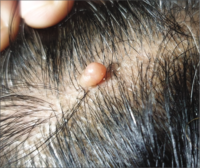
Broad-based pedunculated skin-colored nodular lesion
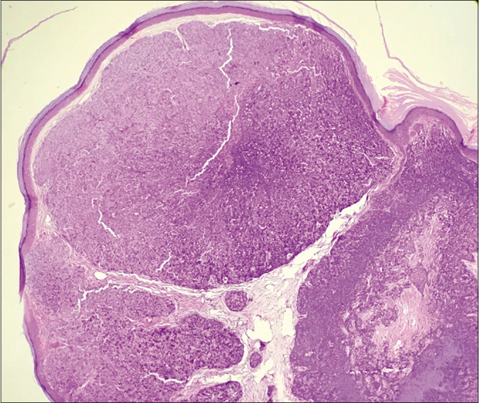
Figure 1:
Broad-based pedunculated skin-colored nodular lesion
Multiple tumor islands with intervening stroma in the dermis (H and E × 40) scanner view
Figure 2:
Multiple tumor islands with intervening stroma in the dermis (H and E × 40) scanner view
Admixture of basaloid cells (green arrows) and scattered mature sebocytes (red arrows) within tumor islands (H and E ×400)
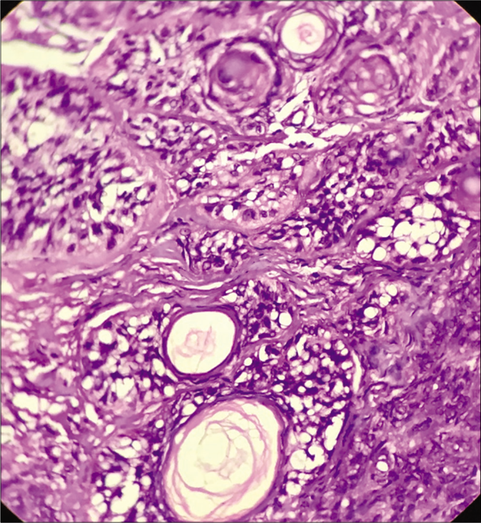
Figure 3:
Admixture of basaloid cells (green arrows) and scattered mature sebocytes (red arrows) within tumor islands (H and E ×400)
Ducts and cystic structures with mature sebocytes (H and E ×400)
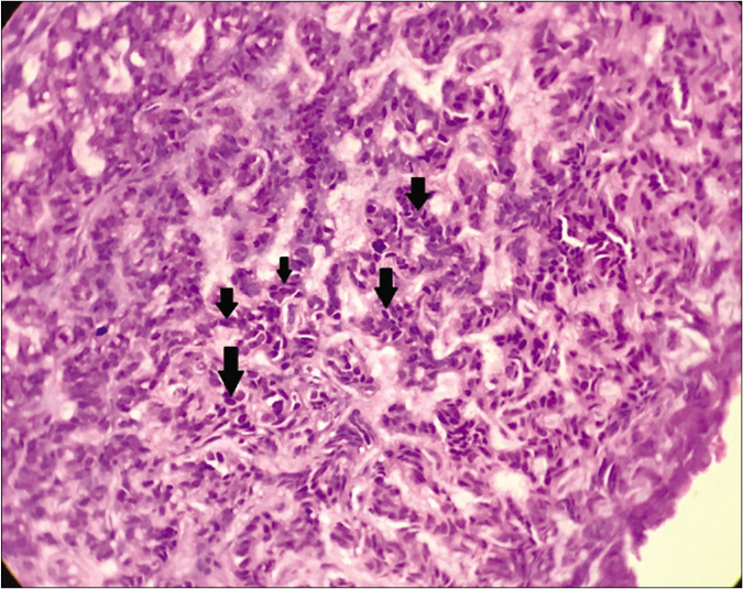
Figure 4:
Ducts and cystic structures with mature sebocytes (H and E ×400)
Labyrinthine/sinusoidal pattern of basaloid cells (black arrows) (H and E ×400)
Figure 5:
Labyrinthine/sinusoidal pattern of basaloid cells (black arrows) (H and E ×400)

Question

What is your diagnosis?

Answer

Sebaceoma

Discussion

Sebaceoma was first described by Troy and Ackerman in 1984 as a benign neoplasm with sebaceous differentiation mostly affecting elderly womenin the head and neck region. It was earlier called sebomatrixoma or sebaceous epithelioma, erroneously indicating malignancy, hence best avoided.1

Classically, these tumors present as yellow or skin-colored nodules of varying sizes either solitary or multiple. They may even occur within the nevus sebaceous. Histologically, the tumor is composed of variable proportions of mature sebocytes and immature sebaceous germinative cells within multiple tumor nodules separated by eosinophilic connective tissue in the dermis. The percentage of immature basaloid cells is usually quite high compared to fewer and mostly scattered mature sebocytes. The connection of tumor nodules with overlying epidermis may or may not be present. Sebaceous ductal structures with holocrine secretions areusually seen within the nodules. Infundibulo cystic structures, areas with apocrine differentiation, squamous metaplasia and keratoacanthoma like architecture are other uncommon features. A few nearly specific organoid patterns which help in the diagnosis of this entity, include rippled pattern, labyrinthine/sinusoidal pattern, carcinoid pattern and petaloid pattern. The rippled pattern is characterized by palisades of neoplastic cells arranged in parallel rows like a ripple, reminiscent of schwannian Verocay bodies. In the labyrinthine or sinusoidal pattern, there is an intricate and tortuous arrangement of various strands and cords of neoplastic cells. Carcinoid pattern comprises trabeculae, ribbons, rosettes and pseudo-rosette-like arrangement of basaloid cells similar to growth patterns in carcinoid tumors. The petaloid pattern shows a flower-like pattern of cells.2

Our provisional clinical diagnosis was cutaneous adnexal neoplasm. Based on clinical features in our case, the important histological differential diagnoses were trichoblastoma or apocrine poroma with sebaceous differentiation. Trichoblastoma with sebaceous differentiation does show large nodular aggregates of basaloid cells and rippled patterns of cells but follicular differentiation and occurrence of germ-papilla arrangement of cells help to differentiate it from sebaceoma. The presence of poroid and cuticular cells with tubular structures and large areas showing apocrine differentiation characterizes apocrine poroma.3

Sebaceous differentiation is usually easily discernible based on mature sebocytes and ducts within the neoplasm. However, in difficult cases, immunohistochemistry marker studies may be done. Mature sebcaeous cells are positive for epithelial membrane antigen and adipophilin.4,5

Dermoscopy serves as an important interface between clinical diagnosis and histological confirmation of skin lesions in current times. Yellowish homogenous ovoid areas correlating with collections of mature sebocytes and arborizing vessels in the periphery of the nodule have been reported which has important clues to the dermoscopic diagnosis of a sebaceoma.6

The occurrence of multiple sebaceous tumors associated with visceral neoplasms, especially gastrointestinal carcinomas characterizes Muir-Torre syndrome which is inherited as an autosomal dominant trait. It is associated with a mutation in genes encoding DNA mismatch repair proteins like MLH1 and MSH2 mainly and less commonly MSH 3, MSH6, MLH3, PMS1 and PMS2.4,5

Herein, we report this case not only for its rarity but also to reinforce the awareness of various histological patterns, differential diagnosis and systemic implications of this unique neoplasm.

Declaration of patient consent

The authors certify that they have obtained all appropriate patient consent.

Financial support and sponsorship

Nil.

Conflicts of interest

There are no conflicts of interest.

References

  1. , , , . Sebaceoma on the scalp simulating a malignant pigmented neoplasia. An Bras Dermatol. 2019;94:590-3.
    [CrossRef] [PubMed] [Google Scholar]
  2. Lesions with predominant sebaceous differentiation In: , , , , eds. Cutaneous Adnexal Teoplasms (1st ed). Philadelphia: Wolters Kluwer Health/Lippincott Williams & Wilkins; . p. :329-408.
    [Google Scholar]
  3. , , , . Sebaceoma and related neoplasms with sebaceous differentiation: A clinicopathologic study of 30 cases. Am J Dermatopathol. 2002;24:294-304.
    [CrossRef] [PubMed] [Google Scholar]
  4. , , , . Sebaceous neoplasia and the Muir-Torre syndrome: Important connections with clinical implications. Histopathology. 2010;56:133-47.
    [CrossRef] [PubMed] [Google Scholar]
  5. , , , , , . Update on the pathology, genetics and somatic landscape of sebaceous tumours. Histopathology. 2020;76:640-9.
    [CrossRef] [PubMed] [Google Scholar]
  6. , , , , . Dermoscopy of rippled pattern sebaceoma. Dermatol Res Pract2010; 2010:140486.
    [CrossRef] [PubMed] [Google Scholar]

Fulltext Views
16,122

PDF downloads
2,971
View/Download PDF
Download Citations
BibTeX
RIS
Show Sections