Translate this page into:
Acquired smooth muscle hamartoma
Correspondence Address:
Sarvesh S Thatte
Department of Dermatology, Seth Gordhandas Sunderdas Medical College and King Edward Memorial Hospital, Parel, Mumbai - 400 012, Maharashtra
India
| How to cite this article: Adulkar SA, Dongre AM, Thatte SS, Khopkar US. Acquired smooth muscle hamartoma . Indian J Dermatol Venereol Leprol 2014;80:483 |
Sir,
A smooth muscle hamartoma is a benign proliferation of smooth muscle bundles within the dermis. [1] Most lesions are congenital and acquired lesions have rarely been reported.
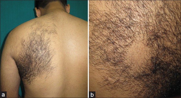
A 32-year-old male presented with increased hair growth on the left side of upper back of 6 months duration. There was no history of itching or pain in the lesion. Cutaneous examination revealed a large patch of hypertrichosis with terminal hair on the left side of upper back in the scapular area [Figure - 1]a. On close observation, the skin in the hypertrichotic area was normal in color. However, on shaving, slightly hyperpigmented small follicular papules were noted [Figure - 1]b.
 |
| Figure 1: (a) A patch of hypertrichosis over the scapular area (b) Follicular papules which were revealed after shaving |
A punch biopsy taken from the hairy area showed mild epidermal hyperplasia with increased basal layer pigmentation. Dermis showed multiple bundles of smooth muscle cells arranged haphazardly in the reticular dermis [Figure - 2]a. On Masson trichrome stain, these bundles stained red in color [Figure - 2]b.
Smooth muscle hamartoma arises from smooth muscle cells that are located in arrector pili muscle, dartos muscle, vascular smooth muscle, muscularis mammillae, and the areolae. [2] Acquired smooth muscle hamartoma is a rare entity. Most of the cases of acquired smooth muscle hamartoma were shown to have originated from arrector pili and dartos muscles. [1] Acquired smooth muscle hamartoma is usually located on the skin of the forearm, chest, neck, scrotum, penis, labia majora, or shoulder.
 |
| Figure 2: (a) Haphazardly arranged multiple bundles of smooth muscle cells in the dermis (H and E, ×100) (b) Smooth muscle bundles stained red (Masson's trichrome, ×200) |
Congenital smooth muscle hamartoma usually manifests at birth, but rarely, can also appear later in life. It is usually located on the trunk and extremities and appears as variably-sized papules, patches, or plaques. [3] Pseudo-Darier′s sign is considered as the characteristic diagnostic clue for congenital smooth muscle hamartoma. [1] Pseudo-Darier′s sign is a transient piloerection and elevation or induration of lesional skin, while Darier′s sign refers to the urtication and erythematous halo which is seen in response to stroking or rubbing of the lesional skin in cutaneous mastocytosis. [4]
In contrast to congenital smooth muscle hamartoma, acquired smooth muscle hamartoma appears later in life as a slightly hyperpigmented or hypertrichotic patch or plaque which may be associated with itching, pain, or numbness. Pseudo-Darier′s sign is usually negative in these lesions. [5]
The histopathologic hallmark of smooth muscle hamartoma is an increase of large, randomly oriented bundles of smooth muscle cells with central, cigar-shaped nuclei. These smooth muscle bundles are arranged haphazardly in the dermis.
Clinically and histologically, Becker′s nevus often resembles smooth muscle hamartoma. Becker′s nevus (also known as Becker′s pigmented hairy nevus) presents as a well defined, uniformly hyperpigmented plaque with hypertrichosis and thickened affected skin that usually manifests at puberty. Histopathology shows epidermal hyperplasia with flattening at the bottom of rete ridges and increased basal layer pigmentation. Occasionally, Becker′s nevus can also show a proliferation of dermal smooth muscles. [6] These clinical and histopathological features help to distinguish Becker′s nevus from acquired smooth muscle hamartoma. Because of the overlapping histopathological features, some authors have speculated that Becker′s nevus and smooth muscle hamartoma are two polar entities at either end of a continuous spectrum. [3]
As acquired smooth muscle hamartoma is a benign condition, no treatment is usually required. However, for cosmetic reasons, laser hair removal and surgical excision may be attempted.
| 1. |
Wong RC, Solomon AR. Acquired dermal smooth-muscle hamartoma. Cutis 1985;35:369-70.
[Google Scholar]
|
| 2. |
Holst VA, Junkins-Hopkins JM, Elenitsas R. Cutaneous smooth muscle neoplasms: Clinical features, histologic finding; and treatment options. J Am Acad Dermatol 2002;46:477-90.
[Google Scholar]
|
| 3. |
Quinn TR, Young RH. Smooth-muscle hamartoma of the tunica dartos of the scrotum: Report of a case. J Cutan Pathol 1997;24:322-6.
[Google Scholar]
|
| 4. |
Surjushe A, Jindal S, Gote P, Saple DG. Darier's sign. Indian J Dermatol Venereol Leprol 2007;73:363-4.
[Google Scholar]
|
| 5. |
Oiso N, Fukai K, Ishii M, Hayashi T, Uda H, Imanishi M. A case of acquired smooth muscle hamartoma of the scrotum. Clin Exp Dermatol 2005;30:523-4.
[Google Scholar]
|
| 6. |
Hsiao GH, Chen JS. Acquired genital smooth-muscle hamartoma: A case report. Am J Dermatopathol 1995;17:67-70.
[Google Scholar]
|
Fulltext Views
4,219
PDF downloads
2,656


