Translate this page into:
Allergic contact dermatitis to Parthenium hysterophorus mimicking actinic prurigo
Correspondence Address:
Sujay Khandpur
Department of Dermatology, Venereology, and Leprology, All India Institute of Medical Sciences, New Delhi - 110 029
India
| How to cite this article: Singh S, Khandpur S, Sharma VK. Allergic contact dermatitis to Parthenium hysterophorus mimicking actinic prurigo. Indian J Dermatol Venereol Leprol 2015;81:82-84 |
Sir,
Allergic contact dermatitis to Parthenium hysterophorus has a wide spectrum of clinical presentations and can mimic various eczematous and photosensitive eruptions. [1],[2] We present a case with an actinic prurigo-like presentation that responded very well to thalidomide.
A 28-year-old unmarried woman presented with a 10-year history of itchy papules and nodules over her extremities and nape of neck. Initially, there were summer exacerbations but then disease became persistent for the last 3 years. She also gave history of episodic, redness of eyes, eyelid pruritus and swelling along with mild facial edema. There was no personal or family history of atopy. Cutaneous examination revealed a predominantly monomorphic, symmetrical eruption of 4-8 mm sized hyperpigmented, dome shaped firm papules involving the nape of the neck and upper back, and the dorsal aspect below knee and elbow, and the ′V′ area of chest. The face, flexures, conjunctiva and oral mucosa were spared. A provisional clinical diagnosis of actinic prurigo was considered.
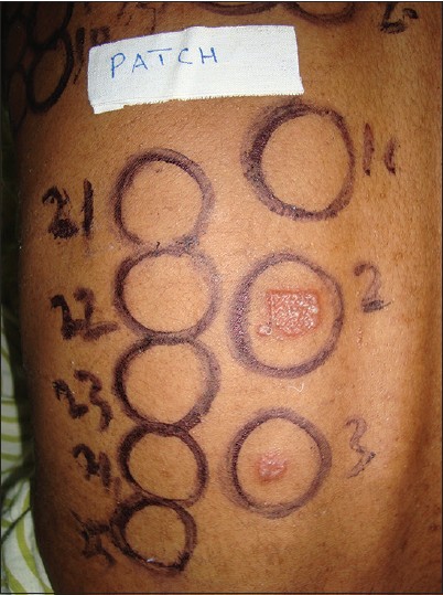
A skin biopsy from a papule showed features of a subacute spongiotic tissue reaction. The baseline hematological and biochemical parameters and anti-nuclear antibody test (ANA) were within normal limits. A patch test using modified Indian Standard Series (ISS) and parthenium antigen showed 3+ positivity to P. hysterophorus aqueous extract in 1:100 dilution [Figure - 1]. Photo-patch testing done using the same antigens with ultraviolet A (UVA) at 10J/cm 2 was negative (2+ positive on both irradiated and non-irradiated sides). Phototesting with narrow band UVB (NbUVB) showed a minimal erythema dose (MED) of 0.8 J/cm 2 and with UVA was negative at 12J/cm 2 .
 |
| Figure 1: Patch test with Indian Standard Series showing 3+ positivity to Parthenium aqueous extract in 1:100 dilution (Patch no.2) and 2+ positivity in 1:200 dilution (Patch no.3) |
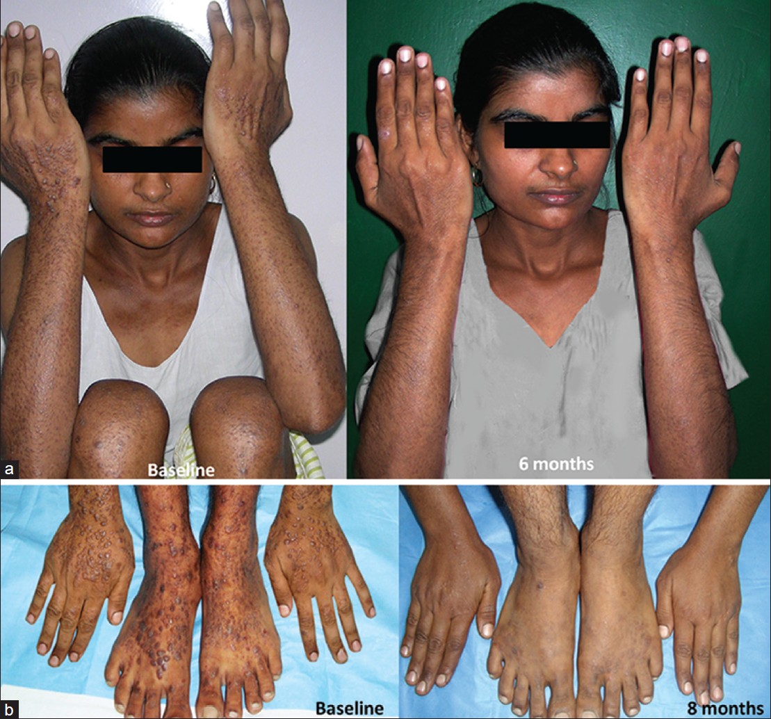
On repeat interviewing, the patient reported lesional exacerbation when visiting her farm to fetch fodder for cattle, especially during summers. She also added that Parthenium was abundant in her surroundings. Hence, a final diagnosis of Parthenium dermatitis presenting with actinic prurigo-like lesions was made. She was started on prednisolone 40 mg/day in tapering doses, but due to minimal response after 3 weeks, it was substituted with thalidomide 100 mg thrice daily after appropriate consent, precautions and counseling. The patient started responding to treatment within 2 weeks and had about 75% improvement after 6 weeks. The lesions flattened completely within 16 weeks and thalidomide was gradually tapered and stopped over the next 6 weeks. It had to be restarted at 100 mg twice daily due to a moderate relapse. The patient had near complete relief with mild scarring and hyperpigmentation [[Figure - 2]a and b]. She was then maintained on thalidomide 100 mg/day for another 10 weeks. The patient did not experience any therapy related adverse effects after 8 months of thalidomide intake. Low dose azathioprine was planned as a maintenance therapy for her but she was lost to follow up.
 |
| Figure 2: Composite clinical photographs showing response to thalidomide. (a) Left side showing baseline image with discrete excoriated papules over extensors of both hands, forearms and 'V' area of chest. Right half showing significant improvement seen after 6 months of thalidomide use. (b) Left side showing baseline image with dome shaped discrete excoriated papules over dorsae of hands and feet. Right half showing near complete clearance after 8 months of thalidomide use |
Parthenium dermatitis in India most commonly presents in a classical airborne contact dermatitis pattern. Other reported presentations include chronic actinic dermatitis, mixed pattern (airborne and photosensitive pattern), exfoliative dermatitis, prurigo nodularis-like eruption and photosensitive lichenoid eruption among others. [1],[2] We were unable to find any previous reports of an actinic prurigo-like presentation due to parthenium dermatitis.
We diagnosed our patient as actinic prurigo in view of the prominent photodistribution of infiltrated, discrete papules and nodules of teenage. In addition, dramatic response to thalidomide has been considered as an indicator of actinic prurigo in the literature. [3] Furthermore, parthenium dermatitis is rare in teenagers and children. [1] However, a strongly positive patch test in the setting of exposure to parthenium in our case suggested parthenium dermatitis. The absence of photopatch test positivity does not refute our clinical diagnosis since parthenium causes enhanced photosensitivity but true photocontact allergy is rare. [1] The MED value in our patient was 800 mJ/cm 2 which is in the normal range for North Indian skin (skin types 3, 4 and 5). [4]
Polymorphous light eruption was not considered as a possibility in the current case as it is characterized by grouped shiny patches, micropapules, papules and plaques on sun-exposed sites. The spongiotic dermatitis on histopathology also favored prurigo over polymorphous light eruption.
Akhtar et al. showed that patients with parthenium dermatitis had significantly elevated levels of TNF-α, IL-6, IL-8 ad IL-17. [5] Thalidomide is known to inhibit TNF-α and IL-8, and also causes alteration of lymphocytic response from Th1 to Th2. [6] These mechanisms may help in other allergic contact dermatitis (ACD) patients as well and suggests a role for thalidomide in cases which are either refractory to systemic glucocorticoids and immunosuppressants or where avoidance of such agents is desired, provided that its benefits outweigh the risk of serious side effects.
| 1. |
Sharma VK, Sethraman G. Parthenium dermatitis. Dermatitis 2007;18:183-90.
[Google Scholar]
|
| 2. |
Verma KK, Sirka CS, Ramam M, Sharma VK. Parthenium dermatitis presenting as photosensitive lichenoid eruption: A new clinical variant. Contact Dermatitis 2002;46:286-9.
[Google Scholar]
|
| 3. |
Ross G, Foley P, Baker C. Actinic prurigo. Photodermatol Photoimmunol Photomed 2008;24:272-5.
[Google Scholar]
|
| 4. |
Tejasvi T, Sharma VK, Kaur J. Determination of minimal erythema dose for narrow band UVB radiation in north Indian patients: Comparison of visual and dermaspectrometer ® readings. Indian J Dermatol Venereol Leprol 2007;73:97-9.
[Google Scholar]
|
| 5. |
Akhtar N, Verma KK, Sharma A. Study of pro- and anti-inflammatory cytokine profile in the patients with parthenim dermatitis. Contact Dermatitis 2010;63:203-8.
[Google Scholar]
|
| 6. |
Wu JJ, Huang DB, Pang KR, Hsu H, Tyring SK. Thalidomide: Dermatological indications, mechanisms of action and side effects. Br J Dermatol 2005;153:254-73.
[Google Scholar]
|
Fulltext Views
3,546
PDF downloads
3,178


