Translate this page into:
An unusual case of multiple angiokeratomas arising on a medium-sized congenital melanocytic nevus
Correspondence Address:
Enzo Errichetti
Via P. Petrone, SNC, 85100 Potenza
Italy
| How to cite this article: Errichetti E, Piccirillo A, Ricciuti F, Ricciuti F. An unusual case of multiple angiokeratomas arising on a medium-sized congenital melanocytic nevus. Indian J Dermatol Venereol Leprol 2012;78:374-375 |
Sir,
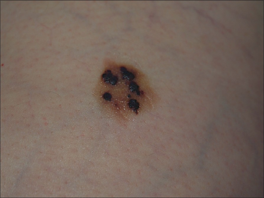
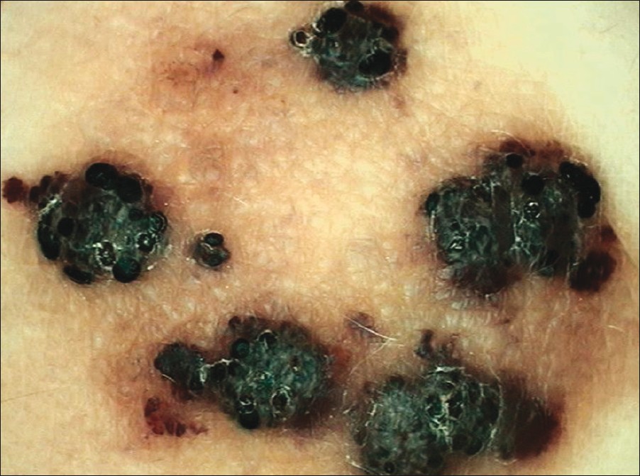
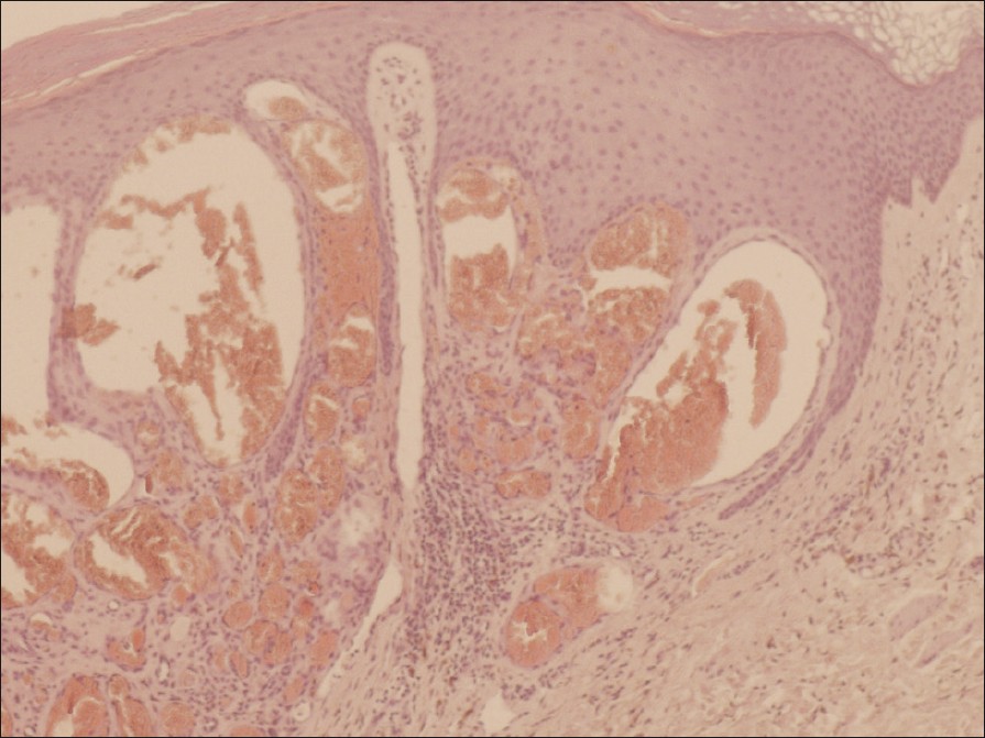
We report the case of a 52-year-old woman, who presented with a light brown plaque (2.2 × 1.5 cm in size) containing multiple, black-violaceous, asymptomatic, keratotic papules on the right thigh [Figure - 1]. The plaque was already present at birth and since then it slowly increased in size until the woman was about 18-year-old, while the first papules appeared when the patient was about 42-year-old. Since then they have gradually increased in number. Dermoscopically, the papules revealed a typical lacunar pattern with sharply demarcated, black lacunas associated with whitish keratotic areas [Figure - 2]. Whereas the light brown plaque revealed a cobblestone pattern [Figure - 2]. Excision of one of the black-violaceous papules on the light-brown plaque was made for histological examination, which showed hyperkeratotic epidermis associated with dilated capillary containing red blood cells in the superficial dermis above nevus cells in the mid-dermis which were distribuited around the dermal blood vessels and between the collagen bundles [Figure - 3]. On the basis of the clinical, anamnestical, dermoscopical and histological data, we made a diagnosis of multiple angiokeratomas arising on a medium-sized congenital melanocytic nevus. The plaque containing papules was completely excised after a patient′s cosmetic request. Congenital melanocytic nevi (CMN) are defined as pigmented skin lesions composed of nevus cells in the epidermis or dermis that are present at birth. [1] They arise from abnormalities of neural crest or its derivatives. [2] The most common classification divides CMN into small (<1.5 cm), medium (1.5-19.9 cm), and large (≥20 cm) based on maximum diameter. Small and medium-sized CMN are primarily a cosmetic concern. In fact, the risk of malignant degeneration of these nevi is low. [1] While malignant neuroectodermal tumors and malignant melanoma may develop more frequently in the lesion of a large-sized CMN. [1],[3] Generally, CMN are infrequently associated with other pathological findings. However, large-sized CMN have been described in association with several conditions such as neurocutaneous melanosis, neurofibromas, lipomas, von-Recklinghausens disease, vitiligo, structural brain malformations, hypertrophy of skull bones and skeletal asymmetry. [3] Anyhow, we are not aware of any previous report of an association between medium-sized congenital melanocytic nevus or other type of congenital melanocytic nevus and multiple angiokeratomas. Furthermore, to the best of our knowledge, there have been no reports of multiple angiokeratomas on a noncongenital melanocytic nevus. Angiokeratomas are benign vascular lesions characterized by ectasia of the superficial dermal vessels and hyperkeratosis of the overlying epidermis. The epidermal changes are secondary to the capillary ectasia. [4] The etiopathogenesis of angiokeratomas remains unknown, but several factors have been involved like increased venous blood pressure or primary degeneration of vascular elastic tissue. [5] In our case, the underlying causes of the association between medium-sized congenital melanocytic nevus and multiple angiokeratomas are unknown. In a report, some authors have explained the co-existence of congenital giant congenital melanocytic nevus, multiple satellite lesions, vitiligo and lipoma in a 17-year-old girl on the basis of a defect in the neural crest, which is considered to be a common origin of melanoblasts, Schwann cells, sensory ganglia, bone, fat, muscle and blood vessels. In particular, it has been suggested that such a defect could lead to abnormal development of any of its derivatives. [2] However, this explicative model is hardly applicable in our case because the angiokeratomas occurred only in adulthood. Anyhow, although the underlying causes of above-mentioned association are unclear, this case is particularly interesting due to its rarity and novelty.
 |
| Figure 1: Light brown plaque (2.2 × 1.5 cm in size) containing multiple, black - violaceous, keratotic papules on the right thigh |
 |
| Figure 2: Dermoscopic image (magnification ×10): The plaque exhibits a cobblestone pattern, while the papules show black lacunas and whitish keratotic areas |
 |
| Figure 3: Hyperkeratotic epidermis associated with dilated capillary containing red blood cells in the superficial dermis (angiokeratoma component) above nevus cells in the mid - dermis which are distributed around the dermal blood vessels and between the collagen bundles (congenital nevus component) (H and E, ×100) |
| 1. |
Conejo-Mir JS, Serrano A, Rodriguez-Freire L, Hernandez C. Pigmented lesions. In: Rusciani L, Robins P, editors. Textbook of Dermatologic Surgery. 1 st ed. Padova: Piccin Nuova Libraria; 2008. p. 379-80.
[Google Scholar]
|
| 2. |
Gulati R, Jain D, Mehrania K, Kuldeep CM, Mathur D. Giant congenital nevomelanocytic nevus with satellite lesions, vitiligo and lipoma: A rare association. Indian J Dermatol Venereol Leprol 2000;66:316-7.
[Google Scholar]
|
| 3. |
Bhagwat PV, Tophakhane RS, Shashikumar BM, Noronha TM, Naidu V. Giant congenital melanocytic nevus (bathing trunk nevus) associated with lipoma and neurofibroma: Report of two cases. Indian J Dermatol Venereol Leprol 2009;75:495-8.
[Google Scholar]
|
| 4. |
Schiller PI, Itin PH. Angiokeratomas: An update. Dermatology 1996;193:275-82.
[Google Scholar]
|
| 5. |
Leis-Dosil VM, Alijo-Serrano F, Aviles-Izquierdo JA, Lazaro-Ochaita P, Lecona-Echeverria M. Angiokeratoma of the glans penis: Clinical, histopathological and dermoscopic correlation. Dermatol Online J 2007;13:19.
[Google Scholar]
|
Fulltext Views
2,904
PDF downloads
1,622


