Translate this page into:
An unusual diagnosis for sporotrichoid nodular lesions: Subcutaneous sarcoidosis
Corresponding author: Dr. Meryem Aktas, Department of Dermatology, Marmara University, Fevzi Cakmak Mah. Muhsin Yazicioglu Cd. No:10, Pendik, Istanbul, Turkey. meryemaktas72@gmail.com
-
Received: ,
Accepted: ,
How to cite this article: Aktaş M, Tunc H, Zaben B, Arı N, Midi I, Cinel L, et al. An unusual diagnosis for sporotrichoid nodular lesions: Subcutaneous sarcoidosis. Indian J Dermatol Venereol Leprol. 2024;90:351-2. doi: 10.25259/IJDVL_582_2021
Dear Editor,
Sarcoidosis is a multi-system disease characterised by non-caseous granulomatous inflammation of unknown aetiology that can affect any organ, most frequently the lung, lymph nodes and skin.1 Skin can be involved in 20–35% of the cases.2 Sarcoid granulomas are typically non-caseating granulomas separated from the surrounding tissue by sharp demarcation, in which macrophages and epithelial cells in the center are surrounded by a few, if any, lymphocytes in the periphery. As the lesions become chronic, the cellularity decreases, and fibrosis which is irreversible, unlike granulomas, becomes more pronounced.1,3 Subcutaneous sarcoidosis, which constitutes approximately 11% of cutaneous sarcoidosis, is characterised by multiple, asymptomatic or slightly tender firm nodules in the subcutaneous tissue with a diameter of 0.5–2 cm and tends to be located on the extremities.4,5 This variant coexists with systemic sarcoidosis in 1.4–6% of patients and has been reported to be associated with systemic disease.3 Herein, we report a case of subcutaneous sarcoidosis with unusual distribution in the setting of severe systemic involvement.
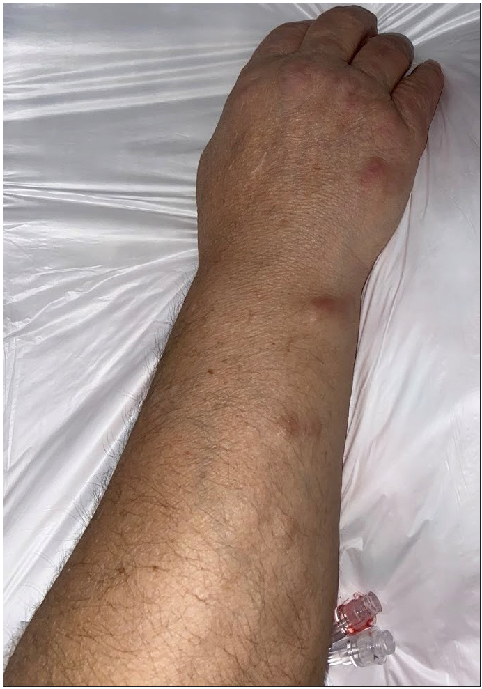
A 55-year-old female patient who presented with erythema nodosum was diagnosed with sarcoidosis after performing thoracic imaging and mediastinoscopic lymph node biopsy in 1994 for respiratory symptoms. Her tuberculin skin and Quantiferon-TB test were negative, and she was in remission without treatment after two years of systemic steroid therapy. In 2007, azathioprine and later methotrexate treatment was given due to the development of muscle weakness related to neurosarcoidosis and eye involvement. Despite five cycles of pulse-cyclophosphamide, neurologic lesions progressed and rituximab treatment was commenced. However, 10 cycles of rituximab treatment, the last one in September 2020, was unable to prevent the emergence of retinal detachment as a consequence of sarcoidosis. She was consulted for skin lesions, when she was hospitalised in March 2021 for pulse-steroid therapy due to eye involvement. Serum calcium, erythrocyte sedimentation rate and C-reactive protein levels were within normal limits, and retest for tuberculin skin test (3 mm) and thoracic imaging for pulmonary infiltration were negative as well. Skin lesions had a history of more than 10 years and changed during treatment, becoming purplish by growing throughout the treatment intervals and shrinking to 3–4 mm with the above-mentioned pulse-cyclophosphamide and rituximab therapies. Physical examination revealed erythematous, firm nodules, 1.5–2 cm in diameter, in a sporotrichoid pattern, on the forearm [Figure 1]. Also, pale erythematous, papules on the dorsum of metacarpophalangeal joints of both hands and an atrophic, desquamated, dark purple patch with a diameter of 2 cm on the anterior surface of the right thigh were noticed. Although the lesions were asymptomatic during steady state, they caused pain when forced between the crutches and the arm during walking. Histopathological examination of an incisional biopsy from a nodule on the forearm revealed prominent fibrosis extending from the deep dermis to the subcutaneous fat tissue and non-caseous granuloma formations trapped in between [Figure 2]. Ziehl–Neelsen and Gomori methenamine silver staining for mycobacterial and fungal infections were negative.

- Erythematous, firm nodules in a sporotrichoid pattern

- Non-necrotizing granulomas in dense fibrotic stroma (hematoxylin and eosin, x100)
Sarcoidosis, denominated as “great imitator,” is characterised by papular lesions, which can mimic rosacea, granuloma annulare, syphilis and lichen planus, while plaque lesions can appear like lupus vulgaris, leishmaniasis, syphilis and psoriasis. Likewise, other clinical presentations of cutaneous sarcoidosis such as alopecia, lupus pernio, hypopigmented plaques, ulcerated lesions and erythroderma can simulate dozens of dermatological diseases including cutaneous lymphomas and vasculitis.2,3 Subcutaneous sarcoidosis should be differentiated from rheumatoid nodules, calcified lipomas and erythema induratum as well.2 Our case was similar to other subcutaneous sarcoidosis cases in the literature in terms of being asymptomatic and erythematous, affecting the extremities and being associated with the systemic disease. However, a thorough search of the existing literature did not reveal a sporotrichoid distribution of subcutaneous sarcoidosis. Subcutaneous sarcoidosis along with superficial venous plexus of forearm has been reported and interpreted to be a reflection of the venous vascular affinity of sarcodosis.6 Although it is well known that sarcoidosis frequently affects the lymphatic system, we do not have enough evidence to associate this lesional distribution with subcutaneous lymphatic involvement.While the diagnosis was easy in our case because of systemic sarcoidosis, we reported the case to emphasize this unusual clinical pattern, an addition to well-known disorders with sporotrichoid pattern; fungal and mycobacterial infections, leishmaniasis, pyogenic lymphocutaneous syndrome, syphilis, metastatic neoplastic lesions and epithelioid sarcomas.7 Thus, dermatologists should be aware of this condition and add diagnostic work-up for sarcoidosis in cases having nodular lesions with sporotrichoid pattern.
Declaration of patient consent
The authors certify that they have obtained all appropriate patient consent.
Financial support and sponsorship
Nil.
Conflicts of interest
There are no conflicts of interest.
References
- Sarcoidosis: A comprehensive review and update for the dermatologist: Part I. Cutaneous disease. J Am Acad Dermatol. 2012;66:699.e1-699.e18.
- [CrossRef] [PubMed] [Google Scholar]
- Cutaneous sarcoidosis: Differential diagnosis. Clin Dermatol. 2007;25:276-87.
- [CrossRef] [PubMed] [Google Scholar]
- Subcutaneous sarcoidosis: Is it a specific subset of cutaneous sarcoidosis frequently associated with systemic disease? J Am Acad Dermatol. 2006;54:55-60.
- [CrossRef] [PubMed] [Google Scholar]
- Subcutaneous sarcoidosis: Report of two cases and review of the literature. Clin Rheumatol. 2011;30:1123-8.
- [CrossRef] [PubMed] [Google Scholar]
- A case of subcutaneous sarcoidosis occurring along the superficial veins of the forearms: A distinctive cutaneous manifestation masquerading venous tropic action in the underlying systemic disease. Case Rep Dermatol. 2017;9:108-13.
- [Google Scholar]
- Lymphocutaneous syndrome. A review of non-sporothrix causes. Medicine (Baltimore). 1999;78:38-63.
- [CrossRef] [PubMed] [Google Scholar]


