Generic selectors
Exact matches only
Search in title
Search in content
Post Type Selectors
Search in posts
Search in pages
Filter by Categories
15th National Conference of the IAOMFP, Chennai, 2006
Abstract
Abstracts from current literature
Acne in India: Guidelines for management - IAA Consensus Document
Addendum
Announcement
Art & Psychiatry
Article
Articles
Association Activities
Association Notes
Award Article
Book Review
Brief Report
Case Analysis
Case Letter
Case Letters
Case Notes
Case Report
Case Reports
Clinical and Laboratory Investigations
Clinical Article
Clinical Studies
Clinical Study
Commentary
Conference Oration
Conference Summary
Continuing Medical Education
Correspondence
Corrigendum
Cosmetic Dermatology
Cosmetology
Current Best Evidence
Current Issue
Current View
Derma Quest
Dermato Surgery
Dermatopathology
Dermatosurgery Specials
Dispensing Pearl
Do you know?
Drug Dialogues
e-IJDVL
Editor Speaks
Editorial
Editorial Remarks
Editorial Report
Editorial Report - 2007
Editorial report for 2004-2005
Errata
Erratum
Focus
Fourth All India Conference Programme
From Our Book Shelf
From the Desk of Chief Editor
General
Get Set for Net
Get set for the net
Guest Article
Guest Editorial
History
How I Manage?
IADVL Announcement
IADVL Announcements
IJDVL Awards
IJDVL AWARDS 2015
IJDVL Awards 2018
IJDVL Awards 2019
IJDVL Awards 2020
IJDVL International Awards 2018
Images in Clinical Practice
Images in Dermatology
In Memorium
Inaugural Address
Index
Knowledge From World Contemporaries
Leprosy Section
Letter in Response to Previous Publication
Letter to Editor
Letter to the Editor
Letter to the Editor - Case Letter
Letter to the Editor - Letter in Response to Published Article
LETTER TO THE EDITOR - LETTERS IN RESPONSE TO PUBLISHED ARTICLES
Letter to the Editor - Observation Letter
Letter to the Editor - Study Letter
Letter to the Editor - Therapy Letter
Letter to the Editor: Articles in Response to Previously Published Articles
Letters in Response to Previous Publication
Letters to the Editor
Letters to the Editor - Letter in Response to Previously Published Articles
Letters to the Editor: Case Letters
Letters to the Editor: Letters in Response to Previously Published Articles
Media and news
Medicolegal Window
Messages
Miscellaneous Letter
Musings
Net Case
Net case report
Net Image
Net Images
Net Letter
Net Quiz
Net Study
New Preparations
News
News & Views
Obituary
Observation Letter
Observation Letters
Oration
Original Article
ORIGINAL CONTRIBUTION
Original Contributions
Pattern of Skin Diseases
Pearls
Pediatric Dermatology
Pediatric Rounds
Perspective
Presedential Address
Presidential Address
Presidents Remarks
Quiz
Recommendations
Regret
Report
Report of chief editor
Report of Hon : Treasurer IADVL
Report of Hon. General Secretary IADVL
Research Methdology
Research Methodology
Resident page
Resident's Page
Resident’s Page
Residents' Corner
Residents' Corner
Residents' Page
Retraction
Review
Review Article
Review Articles
Reviewers 2022
Revision Corner
Self Assessment Programme
SEMINAR
Seminar: Chronic Arsenicosis in India
Seminar: HIV Infection
Short Communication
Short Communications
Short Report
Snippets
Special Article
Specialty Interface
Studies
Study Letter
Study Letters
Supplement-Photoprotection
Supplement-Psoriasis
Symposium - Contact Dermatitis
Symposium - Lasers
Symposium - Pediatric Dermatoses
Symposium - Psoriasis
Symposium - Vesicobullous Disorders
SYMPOSIUM - VITILIGO
Symposium Aesthetic Surgery
Symposium Dermatopathology
Symposium-Hair Disorders
Symposium-Nails Part I
Symposium-Nails-Part II
Systematic Review and Meta-Analysis
Systematic Reviews and Meta-analyses
Systematic Reviews and Meta-analysis
Tables
Technology
Therapeutic Guideline-IADVL
Therapeutic Guidelines
Therapeutic Guidelines - IADVL
Therapeutics
Therapy
Therapy Letter
Therapy Letters
View Point
Viewpoint
What’s new in Dermatology
View/Download PDF

Translate this page into:

Observation Letter
87 (
2
); 259-262
doi:
10.25259/IJDVL_797_19

Anti-neutrophil cytoplasmic antibodies-negative Churg–Strauss syndrome presenting as granuloma annulare-like lesions: An unusual cutaneous presentation and a diagnostic pitfall

Department of Dermatology, The First Affiliated Hospital of Guangzhou Medical University, Guangzhou, P.R. China

Corresponding author: Dr. Weining Huang, Department of Dermatology, The First Affiliated Hospital of Guangzhou Medical University, Guangzhou 510120, P.R. China. 2282233580@qq.com

Licence
This is an open-access article distributed under the terms of the Creative Commons Attribution-Non Commercial-Share Alike 4.0 License, which allows others to remix, tweak, and build upon the work non-commercially, as long as the author is credited and the new creations are licensed under the identical terms.

How to cite this article: Zhu H, Huang M, Huang W. Anti-neutrophil cytoplasmic antibodies-negative Churg–Strauss syndrome presenting as granuloma annulare-like lesions: An unusual cutaneous presentation and a diagnostic pitfall. Indian J Dermatol Venereol Leprol 2021;87:259-62.

Sir,

A 35-year-old male complained of generalized skin rashes. Four years prior to admission to our hospital, the patient had many skin-colored papules on his trunk, limbs and extremities, which were diagnosed as papular granuloma annulare by histological examination at another hospital. The gradual increase in respiratory distress started 6 months prior to admission. It was interpreted as asthma but had no response to inhaled combined steroid therapy and long-acting bronchodilators. Progressively, the patient had visual deterioration and developed tingling with a sensation of numbness of both legs, extending from the posterior thighs to the feet. The patient had a history of sinusitis for more than 10 years and Raynaud’s phenomenon since the previous 7 years.

Dermatological examination showed symmetrical skin-colored and solid papules with a diameter of 0.3–0.5 cm with smooth surface, over his face, neck, trunk, limbs and extremities [Figure 1], without any pustules, ulcers, wheals or purpura. The right upper and lower eyelids were swollen. On respiratory examination, tachypnea and wheezing was noted. Computed tomography scan of the chest showed bronchial thickening, centrilobular nodules, bilateral ground-glass opacity, and tumescent lymph nodes at lung hilus and mediastinum. Sinus computed tomography scan revealed bilateral maxillary, ethmoidal and sphenoidal sinusitis. Ultrasonography showed bilateral cervical lymphadenopathy.
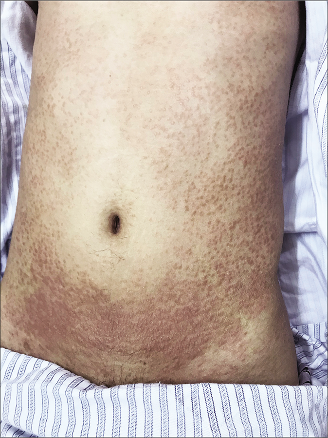
Cutaneous lesions presenting as solid, symmetrical, generalized, skin-colored papules on torso
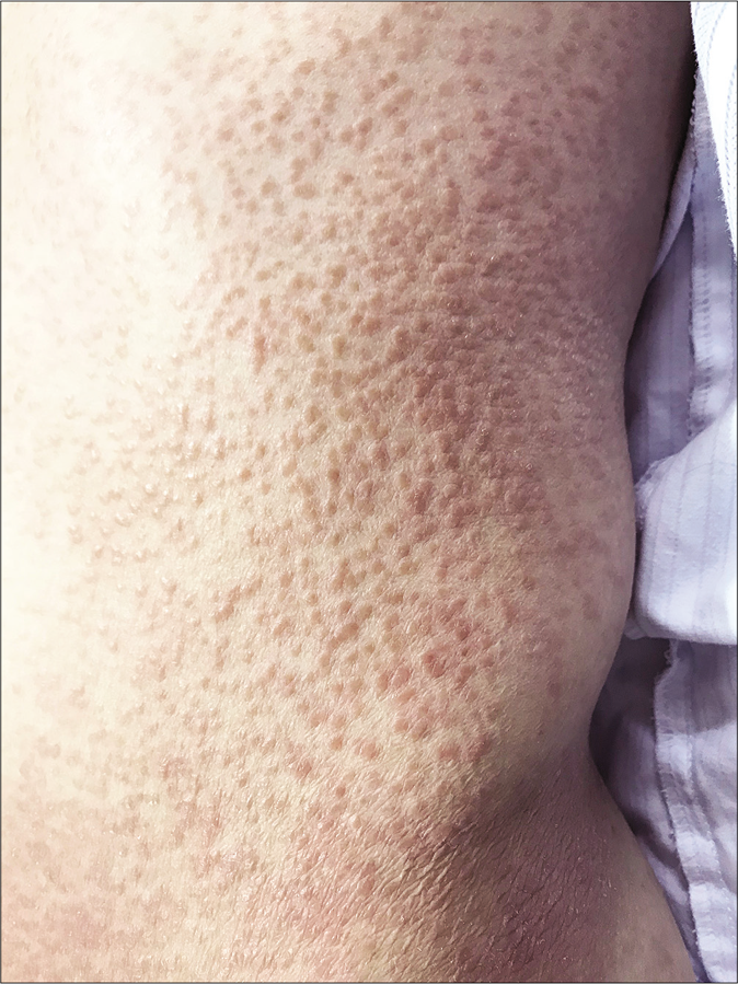
Figure 1a:
Cutaneous lesions presenting as solid, symmetrical, generalized, skin-colored papules on torso
Cutaneous lesions presenting as solid, symmetrical, generalized, skin-colored papules on left flank and trunk
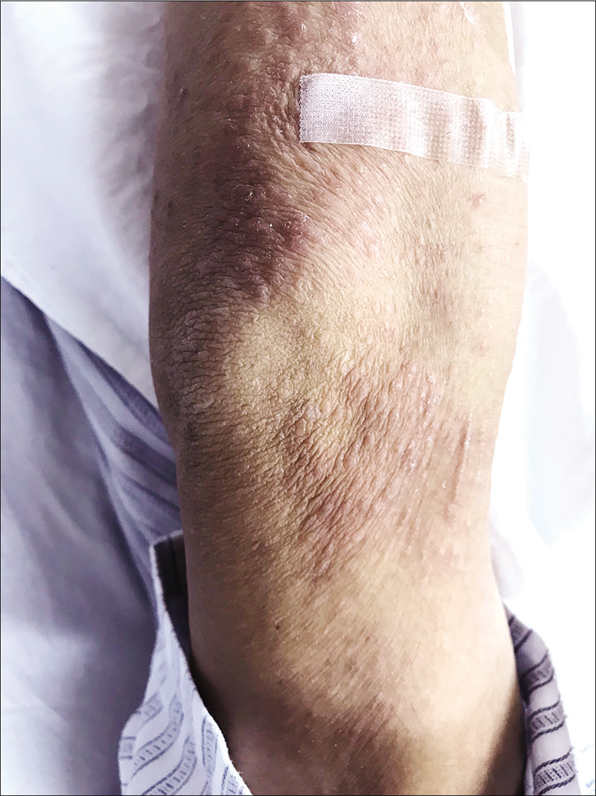
Figure 1b:
Cutaneous lesions presenting as solid, symmetrical, generalized, skin-colored papules on left flank and trunk
Cutaneous lesions presenting as solid, symmetrical, generalized, skin-colored papules on the limb
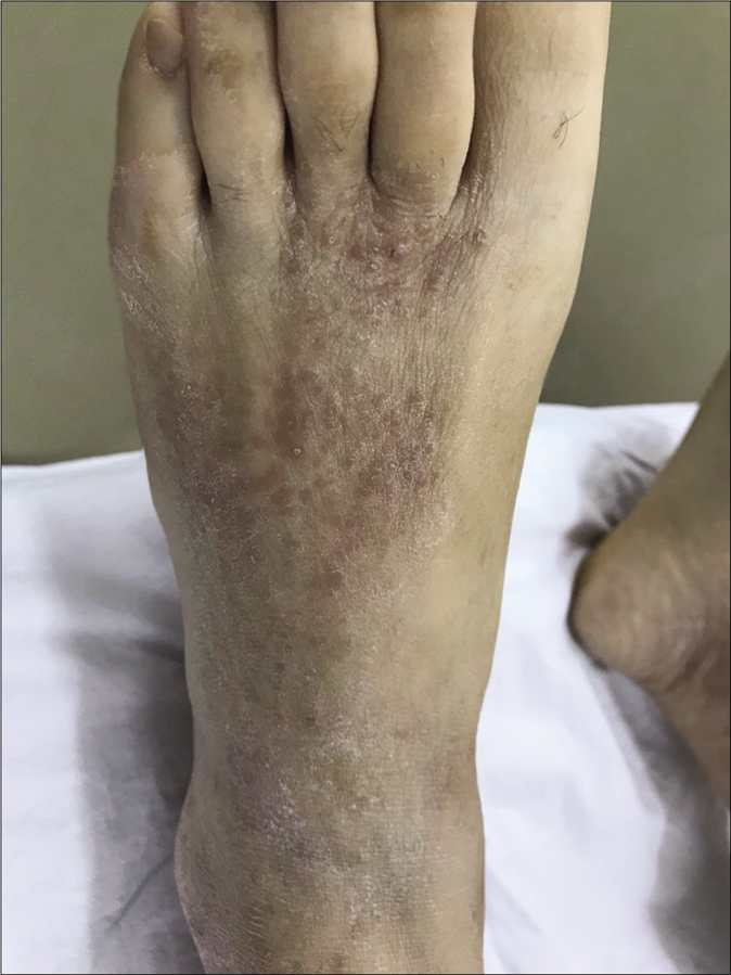
Figure 1c:
Cutaneous lesions presenting as solid, symmetrical, generalized, skin-colored papules on the limb
Cutaneous lesions presenting as solid, symmetrical, generalized, skin-colored papules on left foot
Figure 1d:
Cutaneous lesions presenting as solid, symmetrical, generalized, skin-colored papules on left foot

Complete blood count revealed leukocytosis (24,300/mm3), and marked eosinophilia (15,090 cells/mm3). Elevated immunoglobulin E (716 IU/mL), erythrocyte sedimentation rate (98 mm/h) and C reactive protein level (14.1 mg/dL) were present. Anti-neutrophil cytoplasmic antibodies, antinuclear antibody, and antinuclear antibody profiles were negative. Liver and renal function tests indicated normal levels.

Skin biopsy (hematoxylin-eosin stain) revealed perivascular lymphohistiocytic infiltrate with multinuclear giant cells and eosinophils in the superficial and middermis. There were some lymphohistiocytes scattered between and around the collagen bundles [Figure 2]. Collagen degeneration, dermal mucin, fibrinoid necrosis of the vessel wall, neutrophils or neutrophilic fragments were not observed. Histopathology was reviewed in another hospital and similar findings were reported. Percutaneous lung biopsy revealed diffuse infiltration of lymphohistiocytes and eosinophils in the alveolar septa [Figure 3]. Bone marrow biopsy ruled out any hematologic malignancies.
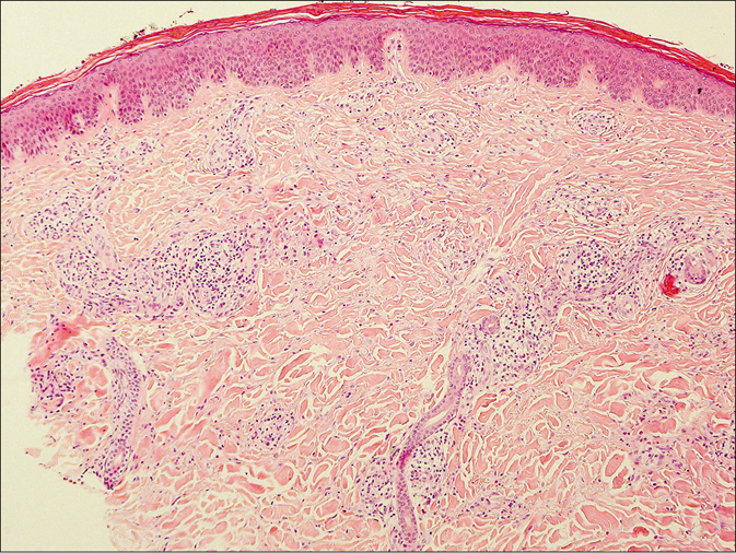
Lymphohistiocytes, multinuclear giant cells and eosinophils infiltrating around vessels and scattered between the collagen bundles (H and E, ×100)
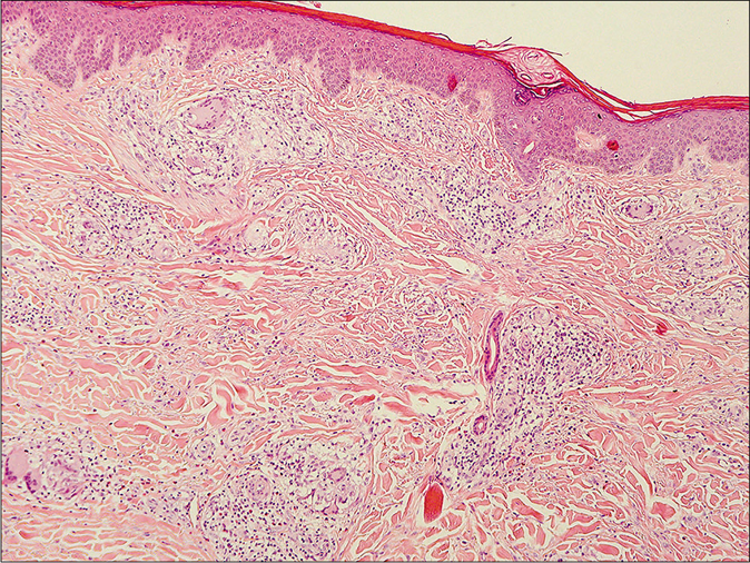
Figure 2a:
Lymphohistiocytes, multinuclear giant cells and eosinophils infiltrating around vessels and scattered between the collagen bundles (H and E, ×100)
Lymphohistiocytes, multinuclear giant cells and eosinophils infiltrating around vessels and scattered between the collagen bundles -- another view (H and E, ×100)
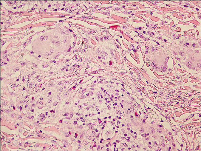
Figure 2b:
Lymphohistiocytes, multinuclear giant cells and eosinophils infiltrating around vessels and scattered between the collagen bundles -- another view (H and E, ×100)
Lymphohistiocytes, multinuclear giant cells and eosinophils infiltrating around vessels and scattered between the collagen bundles - high power view (H and E, ×400)
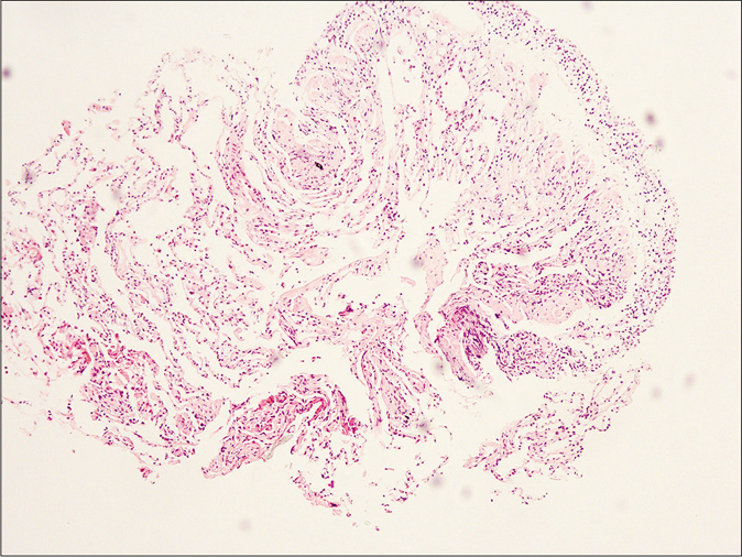
Figure 2c:
Lymphohistiocytes, multinuclear giant cells and eosinophils infiltrating around vessels and scattered between the collagen bundles - high power view (H and E, ×400)
Lymphohistiocytic infiltrate with eosinophils in the alveolar septa (H and E, ×100)
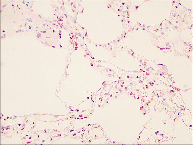
Figure 3a:
Lymphohistiocytic infiltrate with eosinophils in the alveolar septa (H and E, ×100)
Lymphohistiocytic infiltrate with eosinophils in the alveolar septa - high power view(H and E, ×400)
Figure 3b:
Lymphohistiocytic infiltrate with eosinophils in the alveolar septa - high power view(H and E, ×400)

The patient was thus diagnosed with Churg–Strauss syndrome. He was transferred to the respiratory department. Treatment was started with intravenous injection of methylprednisolone (80 mg/d, with decreasing doses) and pulse doses of cyclophosphamide (800 mg once monthly). He displayed gradual improvement in the skin lesions, respiratory distress, tingling sensation and swelling of the eyelids. Unfortunately, he was lost to follow-up 3 months after treatment was initiated.

According to the American College of Rheumatology, Churg–Strauss syndrome can be diagnosed by the presence of any four or more of the following six criteria: asthma, eosinophilia (>10%), neuropathy, migratory pulmonary infiltrates, paranasal sinus abnormalities, and biopsy-proven extravascular eosinophils.1 The patient fulfilled all six diagnostic criteria in our case. This case presented with five clinical manifestations: respiratory distress, sinusitis, polyneuropathy, skin lesions and ocular involvement. Radiologic, laboratory and histological examinations verified five factors: marked eosinophilia, elevated immunoglobulin E, lymphadenopathy, pulmonary infiltration and extravascular eosinophilic infiltration with granulomatous inflammation in the skin lesions. Although orbital involvement is not common, it has been reported in literature. The swelling of the right eyelid was considered as one of the presentations of Churg–Strauss syndrome after exclusion of angioedema, orbital complications secondary to sinusitis and thyroid eye disease.

The typical skin changes in Churg–Strauss syndrome are palpable purpuras, petechiae, ecchymoses, hemorrhagic vesicles and bullae.2 Less frequently, erythematous and purpuric papules with overlying erosions or crusts, confined to the extensor surfaces of the extremities can also be observed.3

We were unable to find any previous reports of cutaneous lesions presenting as symmetrical and generalized papules. In our case, the patient presented with multiple skin-colored papular lesions all over his body, which resembled the cutaneous manifestation of generalized papular granuloma annulare. Isolated and generalized papular presentation without typical cutaneous manifestations in Churg–Strauss syndrome is a rare skin change and may mislead the dermatologist.

The most common pathological characteristics of Churg–Strauss syndrome are extravascular necrotizing granulomas and leukocytoclastic vasculitis.2 There is no single type of vasculitis that appears consistently in such cases. A case presenting with only extravascular granulomas and eosinophils but with the absence of vasculitis has been reported.4 The cutaneous biopsy in our case showed not only perivascular lymphohistiocytes infiltrating with multinuclear giant cells and eosinophils but also interstitial granulomas. On the other hand, the interstitial pattern of histiocytic infiltration is the most common pattern in granuloma annulare, and a number of previous studies have found eosinophils in the cases of this disease.5 However, no collagen degeneration or dermal mucin was observed in our case, which was common in granuloma annulare.5

Eruptive xanthoma, xanthoma disseminatum and Wegener’s granulomatosis were also considered and excluded according to the clinical characters and the results of radiologic, laboratory and histological examination. Isolated and generalized papular presentation is normal in eruptive xanthoma and xanthoma disseminatum however, the histological findings of our case showed extravascular eosinophilic infiltration with interstitial granulomas. No foam cells or extracellular lipid, which are present in eruptive xanthoma and xanthoma disseminatum were abserved. No similar cutaneous manifestations like our case were found in literature in Wegener’s granulomatosis.

Acknowledgment

The authors will thank Professor Baoqing Sun in Guangzhou institute of Respiratory Disease for her great help in this case.

Declaration of patient consent

The authors certify that they have obtained all appropriate patient consent.

Financial support and sponsorship

Nil.

Conflicts of interest

There are no conflicts of interest.

References

  1. , , , , , , et al. The American College of Rheumatology 1990 criteria for the classification of Churg-Strauss syndrome (allergic granulomatosis and angiitis) Arthritis Rheum. 1990;33:1094-100.
    [CrossRef] [PubMed] [Google Scholar]
  2. , , , . Cutaneous manifestations of Churg-Strauss syndrome: A clinicopathologic correlation. J Am Acad Dermatol. 1997;37:199-203.
    [CrossRef] [Google Scholar]
  3. , , , . Cutaneous manifestations of Churg-Strauss syndrome: Key to diagnosis. An Bras Dermatol. 2017;92:56-8.
    [CrossRef] [PubMed] [Google Scholar]
  4. , , , . Granuloma annular-like lesions as a manifestation of eosinophilic granulomatosis with polyangiitis. Rheumatol Clin. 2017;13:357-8.
    [CrossRef] [Google Scholar]
  5. , , , , . Evaluating the unusual histological aspects of granuloma annulare: A study of 30 cases. Indian Dermatol Online J. 2018;9:409-13.
    [Google Scholar]

Fulltext Views
3,937

PDF downloads
4,799
View/Download PDF
Download Citations
BibTeX
RIS
Show Sections