Translate this page into:
Asymptomatic angiomatous lesions on the face and limbs of an adult woman
2 Department of Pathology, Reina Sofía University Hospital, Córdoba, Spain
Correspondence Address:
Carmen Mar�a Alc�ntara-Reifs
Department of Dermatology, Reina Sofía University Hospital, Av. Menéndez Pidal, s/n, 14004 Córdoba
Spain
| How to cite this article: Alc�ntara-Reifs CM, Salido-Vallejo R, Garnacho-Saucedo G, Rizo-Barrios A, Garc�a-Nieto AV. Asymptomatic angiomatous lesions on the face and limbs of an adult woman. Indian J Dermatol Venereol Leprol 2016;82:356-357 |
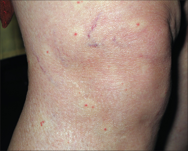
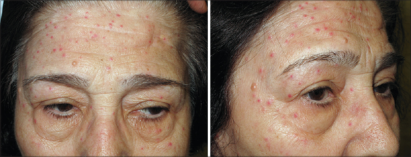
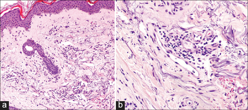
A 73-year-old woman presented with an acute-onset, asymptomatic eruption for 2 days. There was no history of fever or other prodromal symptoms. There was no relevant medical history other than a hepatitis C positivity detected 10 years back for which no treatment was taken. Physical examination revealed erythematous, bright red maculopapular lesions measuring 3–4 mm in diameter on her face [Figure - 1] and limbs. The lesions were blanchable and those on the arms and legs [Figure - 2] were surrounded by pale haloes. Routine laboratory tests including liver function tests were normal. A skin biopsy showed dilated capillaries with plump endothelial cells and a mild lymphocytic infiltrate surrounding the affected vessels in the dermis. The epidermis was unaffected and there was no vascular proliferation [Figure - 3]a and [Figure - 3]b.
 |
| Figure 1: Angiomatous lesions measuring 3–4 mm in diameter on the face |
 |
| Figure 2: Bright red maculopapular lesions surrounded by a peripheral blanched halo on the leg |
 |
| Figure 3: (a) (H and E, ×200) and (b) (H and E, ×400): Dilated dermal blood vessels with plump endothelial cells surrounded by a mild lymphocytic infiltrate |
What Is Your Diagnosis
| 1. |
Higuchi K. About a kind of erythema punctatum. Dermatol Urol 1943;11:171-2.
[Google Scholar]
|
| 2. |
Pérez-Barrio S, Gardeazábal J, Acebo E, Martínez de Lagrán Z, Díaz-Pérez JL. Eruptive pseudoangiomatosis: Study of 7 cases. Actas Dermosifiliogr 2007;98:178-82.
[Google Scholar]
|
| 3. |
Oka K, Ohtaki N, Kasai S, Takayama K, Yokozeki H. Two cases of eruptive pseudoangiomatosis induced by mosquito bites. J Dermatol 2012;39:301-5.
[Google Scholar]
|
| 4. |
Kim JE, Kim BJ, Park HJ, Park YM, Park CJ, Cho SH, et al. Clinicopathologic review of eruptive pseudoangiomatosis in Korean adults: Report of 32 cases. Int J Dermatol 2013;52:41-5.
[Google Scholar]
|
| 5. |
Ban M, Ichiki Y, Kitajima Y. An outbreak of eruptive pseudoangiomatosis-like lesions due to mosquito bites: Erythema punctatum Higuchi. Dermatology 2004;208:356-9.
[Google Scholar]
|
Fulltext Views
3,832
PDF downloads
3,127


