Translate this page into:
Image quality for publication
Correspondence Address:
Feroze Kaliyadan
Department of Dermatology, College of Medicine, King Faisal University, Hofuf
Saudi Arabia
| How to cite this article: Kaliyadan F. Image quality for publication. Indian J Dermatol Venereol Leprol 2016;82:367-370 |
“A picture is worth a thousand words”, goes the famous idiom. However, if an image in dermatology is not of reasonable quality, it may need more than a thousand words to describe! The purpose of this short editorial is to highlight the common reasons why clinical images are deemed unsatisfactory during the editorial process, and how this may result in rejection of the article. Suggestions are provided on how to avoid these common pitfalls.
Some of the common reasons for image rejection are the lack of informed consent, the lesion being out of focus, low resolution, non-centered image, suboptimal image cropping, poor lighting, cluttered background and poorly standardized pre-treatment and post-treatment images.
Informed Consent
Make sure that you have a signed informed consent that specifically mentions the possible use of the image in presentations and publications. Although consent is essential only in images where the facial identity cannot be masked, we suggest taking an explicit consent for photography in all cases.[1] The journal now specifically requires that the consent form should have explicit patient consent not only for photography but also for publication of their images. The consent form should clearly state that the patient has given consent for their images and other clinical information to be reported in the journal, and that they understand that their names and initials will not be published. Although efforts would be made to conceal their identity, anonymity cannot be guaranteed. It is better to make the consent form as explicit as possible.
Center the Image
Unlike artistic photography, clinical photography requires the image to be the center of attention. Frame the image to center the skin lesion or area of interest. Try to fill the frame and avoid too much blank space around the area of interest [Figure - 1]. You can always crop the image to achieve this later but this leads to a loss of pixels thereby leading to a lower resolution image.[1]
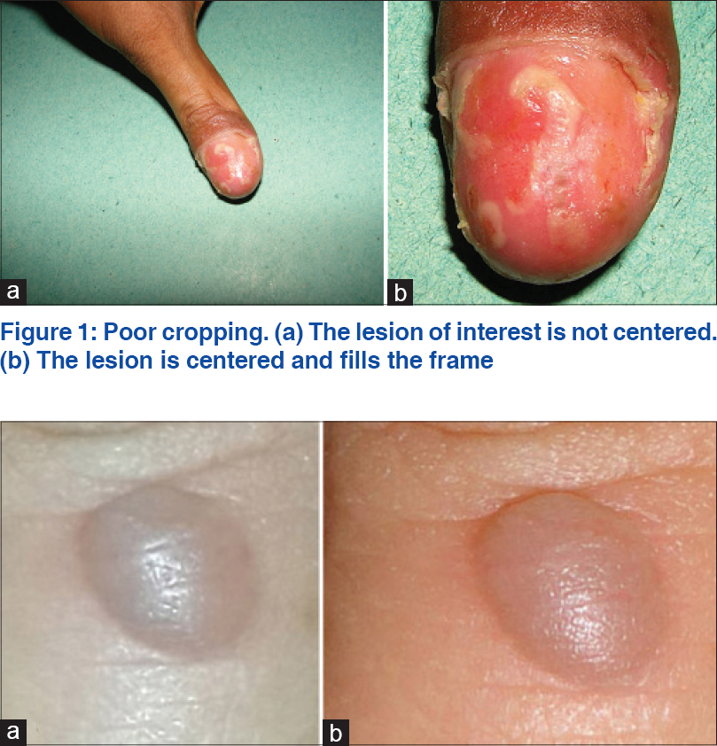 |
| Figure 1: Poor cropping. (a) The lesion of interest is not centered. (b) The lesion is centered and fills the frame |
Use a Good Camera
Smart-phone cameras are quite useful in quickly capturing images for documentation. But most smart-phone cameras come with inherent shortcomings such as a small sensor size and the absence of optical zoom. Image resolution does not depend only on the megapixel capacity of the camera but also the sensor size. Dedicated cameras have a much larger sensor size as compared to smart-phone cameras with the same megapixel capacity. Moreover, most smart-phone cameras do not come bundled with useful add-ons such as a dedicated macro mode (for close-up photography), image stabilization or a good quality flash. The difference becomes more apparent if you have to crop the image [Figure - 2] and [Figure - 3].[2]
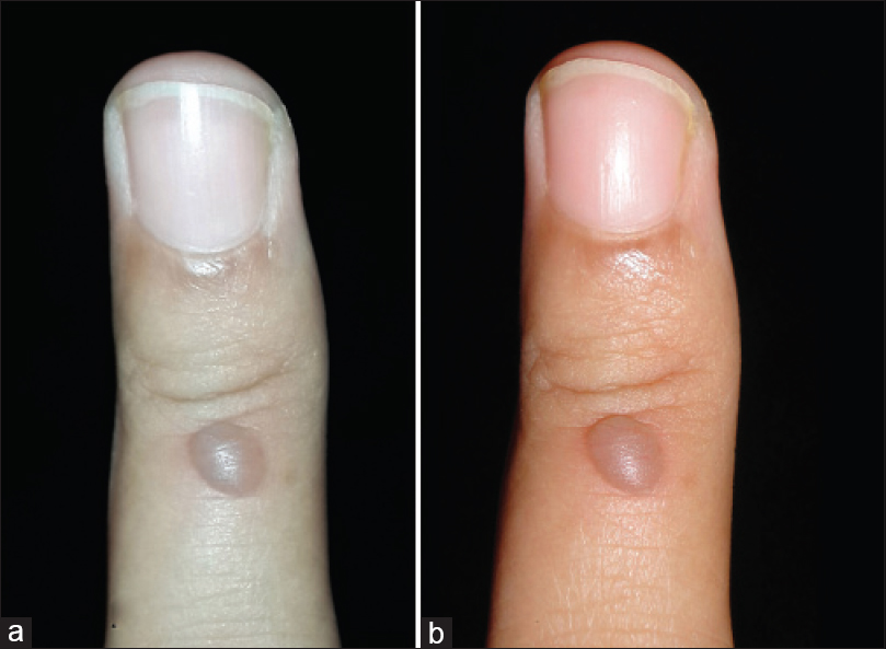 |
| Figure 2: Comparison of cameras. (a) Samsung galaxy note 3 with 13 megapixel resolution (b) Sony cyber shot point and shoot camera with 14.1 megapixel resolution |
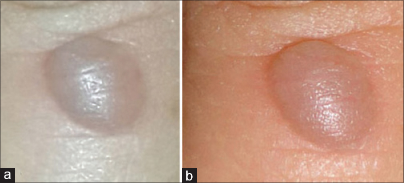 |
| Figure 3: Comparison of cameras (note the further loss of resolution on cropping). (a) Samsung galaxy note 3 with 13 megapixel resolution (b) Sony cyber shot point and shoot camera with 14.1 megapixel resolution |
Today, most point and shoot cameras come with a minimum resolution of 10-megapixels. This is good enough for publication quality images as long as the image does not have to be cropped too much. The usual recommendation for image resolution in print journals is 300 dots per inch. We would also recommend using a camera with a good optical zoom. This helps in photographing the image from a slight distance, and zooming in to capture the lesion. This reduces the optical distortion (seen as a spherical distortion of the area of interest) and also helps to reduce whitening out of the lesion due to a strong flash.
If you do not mind investing a bit more, a digital single-lens reflex would be the way to go. If you publish/present often, or frequently document pre-treatment and post-treatment images, you must seriously consider having a dedicated area and photography set-up in your clinic. Consider investing in equipment such as a tripod, ring flash, dedicated lighting, and macro lenses [Figure - 4].[1],[2],[3]
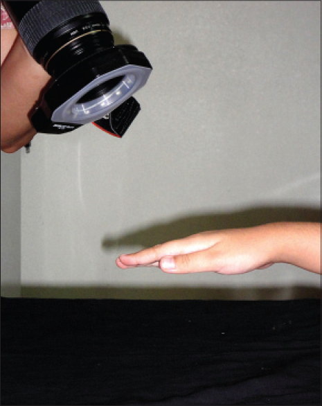 |
| Figure 4: Using a macro lens and ring flash for close-up photography |
Standardize Pre-Treatment and Post-Treatment Images
Good photography is essential for documentation in aesthetic dermatology. Comparative pre-treatment and post-treatment images work best when the images are standardized in terms of lighting and patient position. Various standardization frames are available these days for aesthetic photography. To keep it simple, we suggest using the same camera at the same distance from the patient as in the previous shot. The light source should be the same and the patient should be in the same position.[3]
Lighting and Flash
“Photography” literally means writing with light. Proper lighting is probably the most important factor in any kind of photography. Broad daylight or well-placed overhead lighting of good quality is essential for good clinical photography. With point and shoot cameras, especially when shooting indoors without a tripod, it is better to keep the flash on. This prevents image blurring that happens due to minor camera shakes. However, getting too close to the lesion can lead to a strong reflection from the surface. Overcome this pitfall by shooting from a distance and zooming in. Use good overhead lighting to minimize the use of flash, or even by softening the effect of the flash with a makeshift flash diffuser such as simple butter paper or semi-transparent adhesive tape. When using a digital single-lens reflex camera with a horseshoe flash, you can use reflector boards to diffuse the light [Figure - 5].[1],[3]
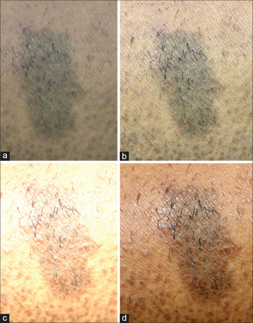 |
| Figure 5: Effect of lighting. (a) Image taken without flash - mild camera shake leading to blurring. (b) Camera stabilized and image taken without flash - minimal movement of the camera or subject can still cause blur. (c) Image with the camera close to lesion - leading to over-exposure and whitening of lesions due to the flash. (d) Image with the camera moved back and zoomed in, with the flash diffused by covering it with a tissue paper |
Background
Use a uniform, non-reflective surface as a background. A cluttered background takes focus away from the area of interest. Most experts suggest using a uniform blue or green colour. However, photographs of some areas such as the nails work best with a dark black background.[1] You can correct the background later using image-editing software such as Photoshop®, but it is always better to do it correctly at the outset itself [Figure - 6].[1]
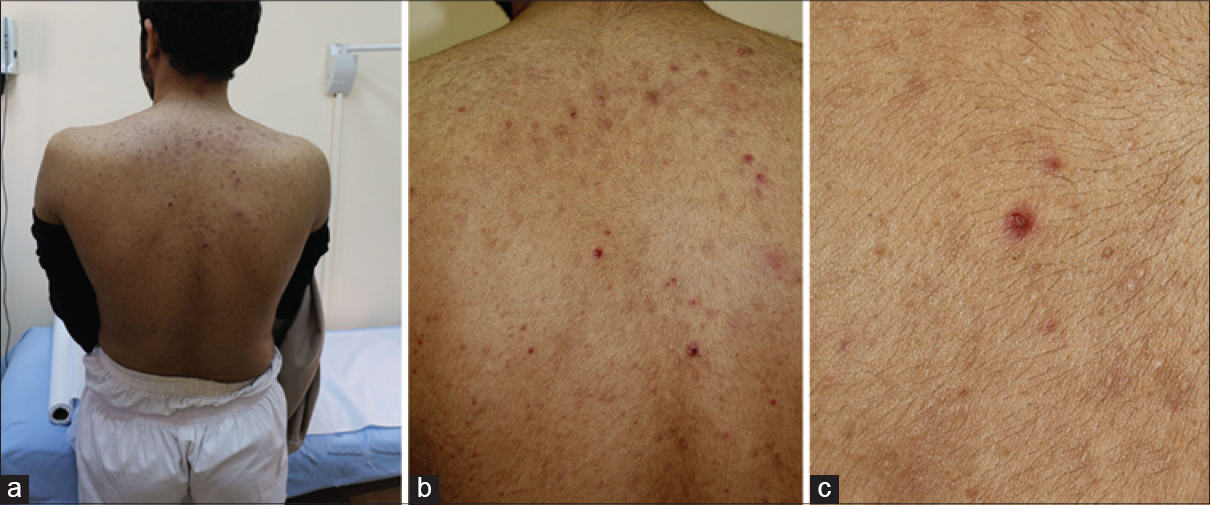 |
| Figure 6: (a) Background distraction. (b) Long shot with a clean background. (c) Close-up shot to show the lesion morphology |
General Tips[1]
- Always take multiple images. Experiment with different angles, distances and lighting conditions. You can always delete the poor images later
- Use the auto-mode of your camera if you are not comfortable with manual settings
- Be familiar with the suggested framing for different types of skin lesions and different areas of the body. For example, in a patient with a generalized rash, take one image from a distance showing the entire area of the rash. Then take a close-up image to highlight the morphology of the individual lesions [Figure - 6]b and [Figure - 6]c. Try to include anatomical landmarks in the frames to give the viewer an idea of the location of the lesion
- Document and catalog your images in a methodical manner. Always make back-up copies
- When submitting images, it is best to submit images as individual files and not as collages. The latter are difficult for the production team to work with
- Editing photographs within ethically acceptable boundaries can help a lot in improving the overall impact of the image. Simple techniques for making collages, clearing background distractions, correcting over-exposure due to flash and enhancing image sharpness or contrast can be easily learned and applied to improve the quality of your images
- Read up on photography in general, get familiar with the basics of photo-editing software and discuss with your colleagues.
| 1. |
Kaliyadan F, Manoj J, Venkitakrishnan S, Dharmaratnam AD. Basic digital photography in dermatology. Indian J Dermatol Venereol Leprol 2008;74:532-6.
[Google Scholar]
|
| 2. |
Ashique KT, Kaliyadan F, Aurangabadkar SJ. Clinical photography in dermatology using smartphones: An overview. Indian Dermatol Online J 2015;6:158-63.
[Google Scholar]
|
| 3. |
Ashique KT, Kaliyadan F. Clinical photography in surgical and esthetic dermatology. In: Mysore V, editor. ACSI (I) Textbook on Cutaneous and Aesthetic Surgery. 1st ed. New Delhi: Jaypee; 2012. p. 883-93.
[Google Scholar]
|
Fulltext Views
3,187
PDF downloads
1,390


![[Figure - 1]](#fig_ijdvl_2016_82_4_367_182968_f1.jpg)
![[Figure - 2]](#fig_ijdvl_2016_82_4_367_182968_f2.jpg)
![[Figure - 3]](#fig_ijdvl_2016_82_4_367_182968_f3.jpg)
![[Figure - 4]](#fig_ijdvl_2016_82_4_367_182968_f4.jpg)
![[Figure - 5]](#fig_ijdvl_2016_82_4_367_182968_f5.jpg)
![[Figure - 6]](#fig_ijdvl_2016_82_4_367_182968_f6.jpg)