Translate this page into:
Becker's nevus among siblings
Correspondence Address:
Varadraj V Pai
Department of Dermatology, Goa Medical College, Goa - 403 202
India
| How to cite this article: Pai VV, Shukla P, Bhobe M. Becker's nevus among siblings. Indian J Dermatol Venereol Leprol 2016;82:359 |
Sir,
Becker's nevus or Becker's melanosis is a relatively common benign condition, which presents as a localized hypermelanosis and hypertrichosis.[1] The familial form of this condition is rare and we were unable to find any previous reports of its occurrence among siblings.
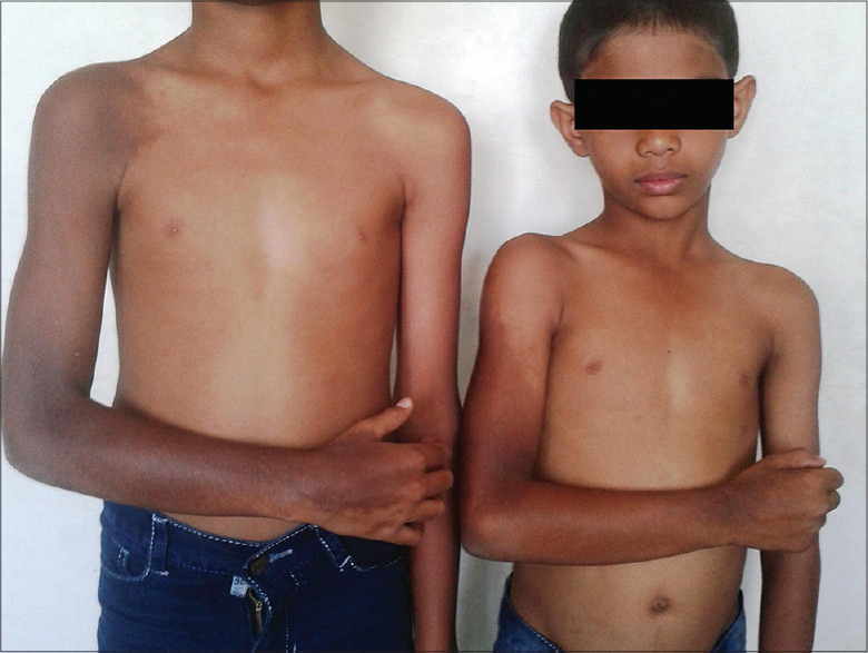
Two brothers, aged 11 and 8 years, born of a non-consanguinous marriage presented aymptomatic pigmented lesions on the upper limb since they were 5 and 2 years old, respectively. Their parents did not have similar lesions. On examination, a hyperpigmented patch was seen extending from the extensor aspect of the arm and forearm till the dorsum of hand in both siblings [Figure - 1]. Involvement of the upper back and scapula with hypertrichosis was noted in the elder sibling. The remainder of the physical examination was normal.
 |
| Figure 1: Becker's nevus extending over the upper extremities of both siblings |
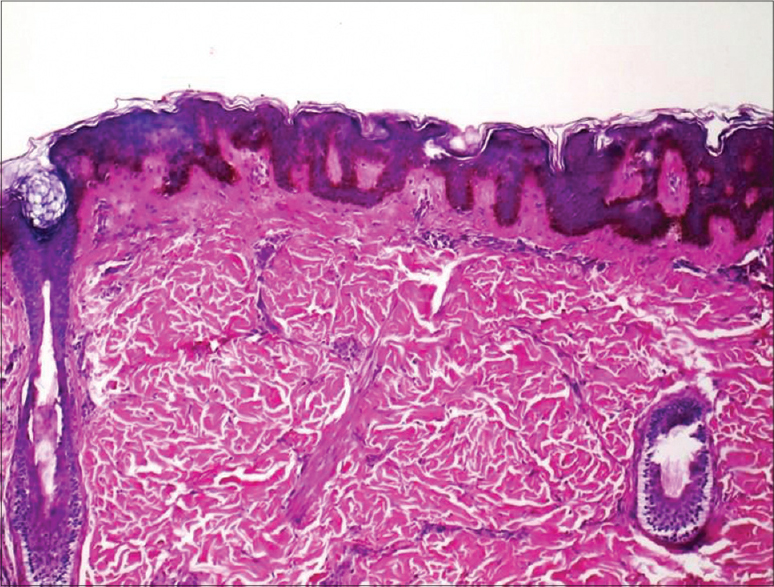
Biopsy revealed epidermal hyperplasia with thickening and elongation of rete ridges with a flat bottom. The basal layer showed increased melanin pigmentation and the dermis revealed a few terminal follicles with arrector pilorum muscles, a picture suggestive of Becker's nevus [Figure - 2].
 |
| Figure 2: Epidermal hyperplasia with thickening and elongation of rete ridges with flat bottoms. The basal layer shows increased melanin pigmentation. The dermis shows a few terminal follicles with arrector pilorum muscles (H and E, ×40) |
Becker's melanosis was described by S. William Becker in 1949 as “concurrent melanosis and hypertrichosis in the distribution of nevus unius lateris”.[1],[2] It is characterized by a well defined hyperpigmented patch with irregular margins and associated hypertrichosis usually over the upper half of trunk and upper extremities.[3]
The prevalence in post-pubertal males is 0.5%. Though the male to female ratio is reported to be 4:1, some workers believe the disease is equally frequent in men and women but cases in women are under-reported and lesions are not conspicuous.[3]
Lesions are usually noticed during adolescence, are initially pale in colour and become more conspicuous after exposure to the sun. It starts as an area of irregular macular pigmentation which spreads to a diameter of several centimetres.[2] The commonest sites are the shoulder, anterior chest and scapular region but lesions on the face, neck and distal limbs have been reported.[4]
Atypical presentation of Becker's nevus can be related to onset (congenital or late), family history, rare site of distribution like legs, presence of multiple lesions, bilateral distribution or dermatomal or blaschkoid pattern of involvement.[3],[5] Though there are some cases reported of familial Becker's nevus, almost all have a parent–child association.[5],[6],[7]
The exact pathogenesis of Becker's nevus is unknown. It may be due to mosacism wherein the lesions follow a blaschkoid, checkerboard, phylloid pattern or a patch without midline separation due to somatic mutation where the abnormal phenotype expression occurs only in the affected segments of the body resulting in a linear or block-like configuration.[8] It is believed that androgens may play a role in Becker's melanosis as evidenced by its peri-pubertal development, male preponderance, hypertrichosis, acneiform lesions within the patch and increase in androgen receptors in involved skin.[2]
Becker nevus syndrome includes the association of Becker's nevus with one or more of the following: unilateral hypoplasia of the breast, aplasia of the ipsilateral pectoralis major muscle, ipsilateral limb shortening, localized lipoatrophy, spina bifida, scoliosis, pectus carinatum, congenital adrenal hyperplasia and an accessory scrotum.[4]
Histopathology shows acanthosis, broad and fused adjacent rete ridges, variable hyperkeratosis and basal layer hyperpigmentation. Associated dermal smooth muscle hyperplasia led some workers to propose that Becker nevus is in a continuum of hamartomatous processes along with congenital smooth muscle hamartoma. Becker melanosis without smooth muscle hyperplasia has been reported.[4]
Treatment includes Q-switched or long pulsed ruby or Nd:Yag laser or Er:Yag laser for hyperpigmentation and hypertrichosis. Cosmetic camouflage is also useful.[4]
Declaration of patient consent
The authors certify that they have obtained all appropriate patient consent forms. In the form the patient(s) has/have given his/her/their consent for his/her/their images and other clinical information to be reported in the journal. The patients understand that their names and initials will not be published and due efforts will be made to conceal their identity, but anonymity cannot be guaranteed.
| 1. |
Becker SW. Concurrent melanosis and hypertrichosis in distribution of nevus unis lateris. Arch Derm Syphilol 1949;60:155–60.
[Google Scholar]
|
| 2. |
Naveen KN, Pai VV, Hegde S, Atnanikar SB, Athanikar V. Becker's naevus on the forearm-A case report. Egypt Dermatol Online J 2013;9:9.
[Google Scholar]
|
| 3. |
Alfadley A, Hainau B, Al Robaee A, Banka N. Becker's melanosis: A report of 12 atypical cases. Int J Dermatol 2005;44:20–4.
[Google Scholar]
|
| 4. |
Moss C, Shahidullah H. Nevi and other developmental defects. In: Burns T, Breathnach S, Cox N, Griffiths C, (editors). Rook's textbook of dermatology. Vol. 18. Oxford: Wiley-Blackwell; 2010. p. 17–9.
[Google Scholar]
|
| 5. |
Fretzin DF, Whitney D. Familial Becker's nevus. J Am Acad Dermatol 1985;12:589–90.
[Google Scholar]
|
| 6. |
Book SE, Glass AT, Laude TA. Congenital Becker's nevus with a familial association. Pediatr Dermatol 1997;14:3735.
[Google Scholar]
|
| 7. |
Panizzon R, Schnyder UW. Familial Becker's nevus. Dermatologica 1988;176:2756.
[Google Scholar]
|
| 8. |
Ro YS, Ko JY. Linear congenital Becker nevus. Cutis 2005;75:122–4.
[Google Scholar]
|
Fulltext Views
4,475
PDF downloads
2,021


