Translate this page into:
Bullous impetigo mimicking epidermolysis bullosa
Corresponding author: Dr. Biswanath Behera, Department of Dermatology. All India Institute of Medical Sciences, Bhubaneswar - 751 019. Odisha, India. biswanathbehera61@gmail.com
-
Received: ,
Accepted: ,
How to cite this article: Dash S, Palit A, Behera B. Bullous impetigo mimicking epidermolysis bullosa. Indian J Dermatol Venereol Leprol 2022;88:851-2.
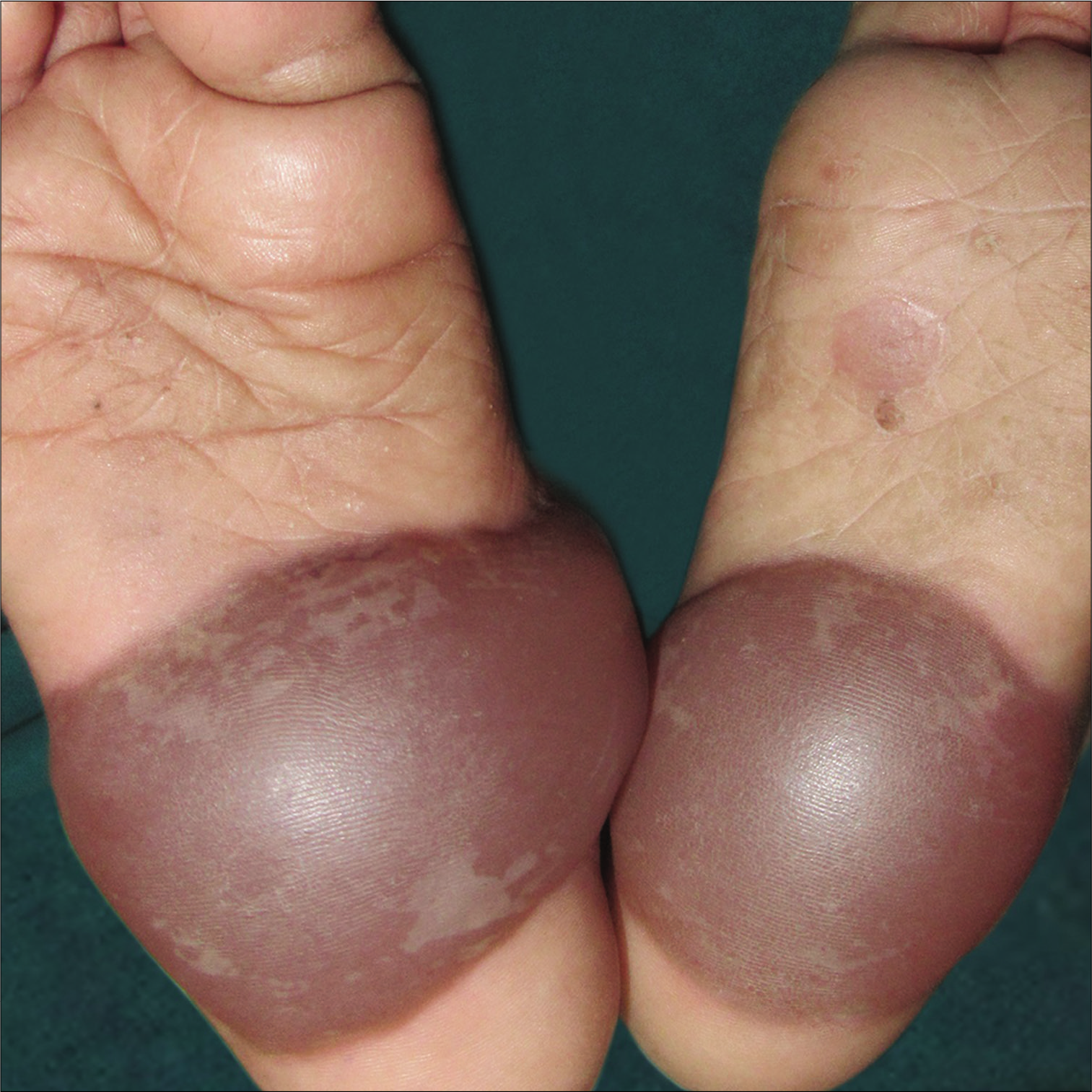
A 2-year-old boy was brought with 4 days history of 2 fluid-filled lesions on soles. It was associated with minimal itching and there were no constitutional symptoms. Cutaneous examination revealed hemorrhagic bullae of approximately 4×5 cm in size on soles [Figure 1]. One healing erosion was present on the left sole. Other cutaneous, mucosal, teeth and nail examinations were within normal limits. Differential diagnosis of bullous impetigo and epidermolysis bullosa was considered. The aspirated fluid was sent for bacterial culture sensitivity and oral amoxicillin with clavulanic acid 375 mg twice daily was started empirically. Aspirated fluid had grown methicillin-resistant Staphylococcus aureus. A final diagnosis of bullous impetigo was made. The lesions subsided completely after 7 days of treatment.

- Hemorrhagic bullae on soles
Declaration of patient consent
The authors certify that they have obtained all appropriate patient consent.
Financial support and sponsorship
Nil.
Conflicts of interest
There are no conflicts of interest.


