Translate this page into:
Clear cell hidradenoma in a young boy
2 Department of Pathology, Ahwaz Jondi Shapour, University of Medical Sciences, Ahwaz, Iran
Correspondence Address:
Reza Yaghoobi
Department of Dermatology, Emam Khomeini Hospital, 61335, Ahwaz
Iran
| How to cite this article: Yaghoobi R, Kheradmand P. Clear cell hidradenoma in a young boy. Indian J Dermatol Venereol Leprol 2007;73:358-359 |
 |
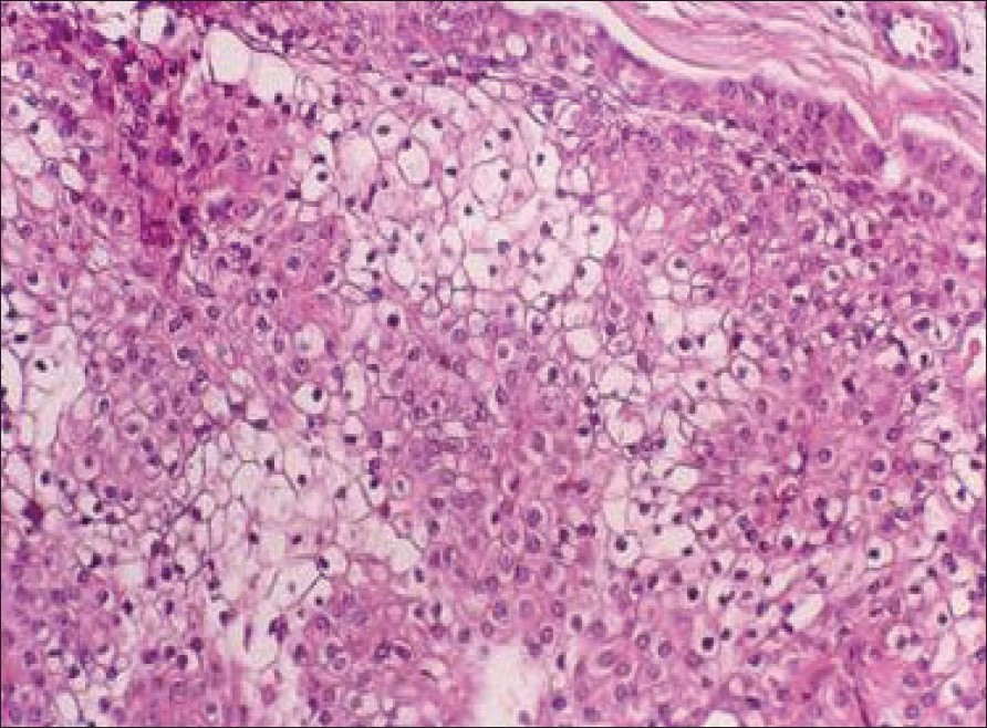
| Figure 2: Histopathology showing clear and pale cells (H and E, X200) |
 |
| Figure 2: Histopathology showing clear and pale cells (H and E, X200) |
 |
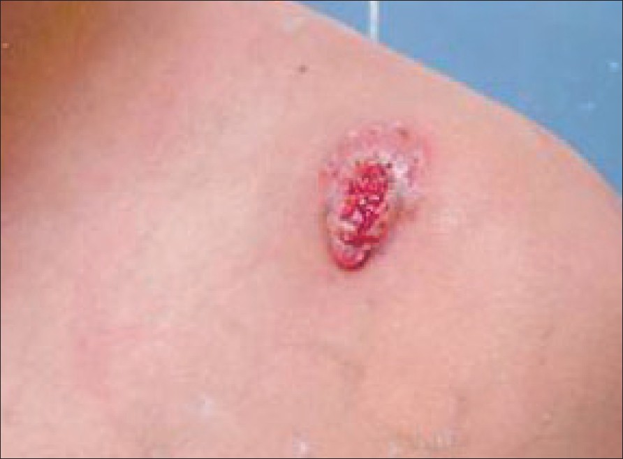
| Figure 1: Solitary tumor with ulcerated surface on the left shoulder |
 |
| Figure 1: Solitary tumor with ulcerated surface on the left shoulder |
Sir,
Clear cell hidradenoma is an uncommon benign tumor differentiating towards sweat glands. These tumors are variable in size with a small tendency to ulcerate and have a low malignant potential. [1] Usually, these are diagnosed between the fourth and the eighth decade of life, with a peak incidence in the sixth decade. [2] Women are affected more often than men. [3] We present the case of a 14-year-old boy with an ulcerated clear cell hidradenoma.
A 14-year-old boy presented with a progressively enlarging nodule on his left shoulder for the past 1 year. There was no preceding history of trauma involving the affected area. Clinical examination revealed a 1.0 x 0.5cm, firm, nontender, erythematous nodule on the left shoulder with an ulcerated surface [Figure - 1]. There was no regional lymphadenopathy. The boy was otherwise in good health.
The tumor was excised. Histopathological examination revealed a circumscribed nonencapsulated multilobulated tumor centered in the dermis with epidermal connections. The tumor was composed of solid nests of round cells with eosinophilic or clear cytoplasm. There were a few duct-like structures lined by one-layered cuboidal cells [Figure - 2] that were suggestive of clear cell hidradenoma. The postoperative course was uneventful as observed during the follow-up after 3 months.
Mayer first described hidradenoma as a distinct clinical entity. [4] These are benign adnexal neoplasms of uncertain origin. Majority of the investigators consider these neoplasms to be of eccrine origin. However, there are occasional reports supporting an apocrine derivation. Recently, hidradenoma has been classified into two groups; those with eccrine differentiation (poroid hidradenoma) and those exhibiting apocrine differentiation (clear cell hidradenoma). [4] Clear cell hidradenomas are by far the most frequent subtype and account for 95% of all the cases. [2],[5]
Clinically, clear cell hidradenoma presents as a slow growing, solitary, freely mobile and firm nodule. Occasionally, there may be a cystic appearance. The lesions may be skin-colored, red, blue or brown; multiple lesions are rare. There is a predilection for occurrence over the scalp, face, thorax and abdomen; however, the tumor may be located anywhere on the body. [3] Rarely, malignant transformation of clear cell hidradenomas with metastasis has been reported. [2] The presentation of our patient is considered to be unusual because of the early age of onset and clinical picture.
Clear cell hidradenoma is well circumscribed and is sometimes encapsulated. It may be connected to the epidermis and the dermal epithelial lobules may extend into the subcutaneous fat. Tubular lumina of varying sizes are often present within the lobulated masses. These may bifurcate and are usually lined by the cuboidal epithelium and sometimes show glandular structures surrounded by columnar cells. Although it may contain a mixture of solid and cystic areas in varying proportions, clear cell hidradenoma is considered to be a solid tumor. The main histopathologic characteristic of clear cell hidradenoma is the presence of large pale to clear cells with small, monomorphous, round or plump oval and often eccentrically located nuclei. The clear cells contain considerable amount of glycogen; however, in addition, they may show significant amount of PAS - positive, diastase - resistant material along their periphery. [2],[5]
The diagnosis of clear cell hidradenoma solely on the basis of the clinical appearance of the lesion is difficult. The sites of occurrence are nonspecific and similar to other nodulocystic lesions of the skin. The histopathological characteristics of clear cell hidradenoma are distinctive, but the lesion should be differentiated from other conditions where clear cells may be present. Surgical removal with wide margins is recommended for this tumor as there is a high rate of local recurrence and this tumor has the potential for malignant transformation. [3] A review of a large series of patients with clear cell hidradenoma has shown a postsurgical recurrence rate of approximately 10%. This may be attributed to the incomplete resection and the fact that the tumor tissue insinuates through the dermis and subcutaneous tissue. [5]
| 1. |
Feldman AH, Niemi WJ, Blume PA, Chaney DM. Clear cell hidradenoma of the second digit: A review of the literature with case presentation. J Foot Ankle Surg 1996;36:21-3.
[Google Scholar]
|
| 2. |
Faulhaber D, Worle B, Trautner B, Sander CA. Clear cell hidradenoma in a young girl. Am Acad Dermatol 2000;42:693-5.
[Google Scholar]
|
| 3. |
Schweitzer WJ, Goldin HM, Bronson DM, Brody PE. Ulcerated tumor on the scalp. Clear-cell hidradenoma. Arch Dermatol 1989;125:985-6.
[Google Scholar]
|
| 4. |
Gianotti R, Alessi E. Clear cell hidradenoma associated with the Folliculo - Sebaceous - Apocrine unit: Histologic study of five cases. Am J Dermatopathol 1997;19:351-7.
[Google Scholar]
|
| 5. |
Ozawa T, Fujiwara M, Nose K, Muraoka M. Clear cell hidradenoma of the Forearm in a young boy. Pedia Dermatol 2005;22:450-2.
[Google Scholar]
|
Fulltext Views
2,826
PDF downloads
1,525


