Translate this page into:
Comparative histopathology of scabies versus nodular scabies
Correspondence Address:
R R Mittal
Departments of Dermato-Venereology and Pathology, Government Medical College/Rajindra Hospital, Patiala - 147 001
India
| How to cite this article: Mittal R R, Singh S P, Dutt R, Gupta S, Seth P S. Comparative histopathology of scabies versus nodular scabies. Indian J Dermatol Venereol Leprol 1997;63:170-172 |
Abstract
Comparative histopathology was studied in 25 cases of scabies versus 25 cases of nodular scabies which were selected from Dermato-Venereology out patients. Salient differences observed were that in scabies lifting of stratum corneum at places was seen in all 100% cases, spongiosis in 100%, spongiotic vesicles in 28%, burrows in 56%, mite in 40% and vasculitis in 28% whereas in nodular scabies acanthosis was seen in 100%, pseudo epitheliomatous hyperplasia in 8%, burrows in 48%, mite in 24% and vasculitis in 84%. In nodular scabies, dermal infiltrate in 32% cases was arranged as lymphoid follicles with admixture of plasma cells and eosinophils. |
 |
 |
 |
Scabies (S), a contagious parasitic disorder is caused by itch mite, Sarcoptes scabiei. Histopathologically, papular lesions of S reveal burrows with mite, empty burrows, eggs containing larvae, egg shells or excreta.[1] Faecal material or scybala within the horny layer is also indicative of scabies.[2] Spongiosis and spongiotic vesicles are seen in stratum malpighii near the burrows in S.[2] Vasculitis and dermal infiltrate in sections containing mite in S show variable number of eosinophils.[3] Mites are hardly ever, found in nodular scabies (NS). Histopathologically, nodules reveal dense chronic inflammatory infiltrate resembling lymphoma with atypical mononuclear cells.[4] Eosinophils may be present[1] or absent in lymphoma-like infiltrate.[3] Vasculitits with thickened walls, fibrinoid deposits, and vessel wall infiltration by cells was seen in NS.[1]
Subjects and Methods
Twenty-five cases of S and 25 cases of NS diagnosed clinically were selected from Dermato-Venereology outpatients for this study. Detailed history, thorough clinical and dermatological examination were done. Lesions were cleaned with spirit and infiltrated with 2% xylocaine. Elliptical biopsy was taken with the help of a scalpel. Samples were preserved in 10% formalin and sent for histopathology. Haematoxylin and eosin staining was done and slides were observed under 1OOx and 400x magnification with the help of light microscope.
Results
Hyperkeratosis was observed in 92% cases of S and in all cases of NS. Follicular keratosis was seen in all cases of S compared to 16% cases of NS. Delling was seen in all cases of S but in only 12% of NS. Lifting of stratum corneum was seen in 100% cases of S and it was absent in NS. Mild to moderate acanthosis was observed in 88% cases of S and more pronounced acanthosis in 100% of NS. In 8% cases of NS, pseudoepitheliomatous hyperplasia was seen.
Burrows were seen in 56% cases of S, in 3 of them they traversed full strartum malpighii and 10 of them had mite or scybala. In NS, 48% showed burrows and 6 of them had mite or scybala. In S, spongiosis was seen in all cases, more prominent near burrows and with spongiotic vesicle formation in 7/25 (28%) cases, and bullae in one case. [Figure - 1] In NS, milder spongiosis was seen in 14, homogenisation of epidermal cells in 3 and intracellular oedema in 5 cases.
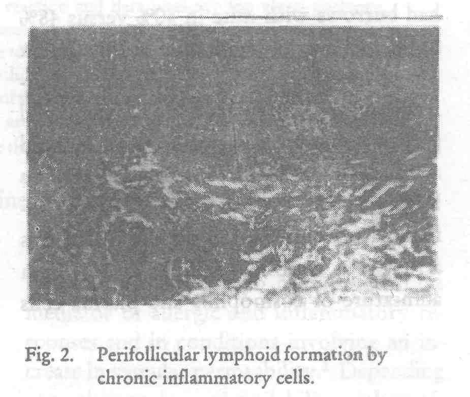
Papillomatosis was seen in 16 cases of NS versus 8 of S. Oedema of dermis was seen in 100% cases of S versus 80% of NS. Perivascular mononuclear infiltrate was seen in all 50 cases. Infiltrate was mild in S and moderate to intense in NS. In 10/25 cases of NS, prominent periadenexal infiltrate was also seen. In 8/25 cases of NS, infiltrate revealed lymphoid follicle formation with paler cells in the centres and darker cells towards periphery. Atypia and mitotic cells were seen in 48%; eosinophils in 72% and the plasma cells in 48% cases of NS, and these cells were observed even where infiltrate was not arranged as lymphoid follicles. Vasculitis seen as thickened vessels due to multilayering of endothelial cells, fibrinoid depostis and cellular infiltration of vessel walls was seen in 28% cases of S verus 84% of NS. Fibrinoid deposits in vessel walls was more in S and irregular thickening due to cellular poliferation was more in NS.
Discussion
Salient histopathological difference in S versus NS were (i) lifting of stratum corneum in 100% of S and its absence in NS (ii) more pronounced spongiosis and spongiotic vesicle formation in S versus more of intracellular oedema and homogenisation of epidermal cells in NS (iii) Fifty-six percent cases of S had burrows with mite in 40% versus 48% of NS had burrows with mite in 24% (iv) six out of 25 cases of NS had mite in the burrows and therefore persistent antigen could play important role in pathogenesis. Marked acanthosis in 100% with pseudoepitheliomatous hyperplasia in 8% moderate to intense inflammatory infiltrate in dermis which was arranged as lymphoid follicles in 32% with admixture of eosinophils and plasma cells were characteristic of NS and all above features differntiated lymphoid follicles of NS from lymphomas. Vasculitis was seen in 28% of cases of S versus 84% of NS.
| 1. |
Fernandez N, Torres A, Ackerman A B. Pathological findings in human scabies, Arch Dermatol 1977;113:320-324.
[Google Scholar]
|
| 2. |
Lever W F, Lever G S. Histopathology of scabies, in: Histopathology of the Skin, 7th edition J B Lippin cott Company, 1990;238.
[Google Scholar]
|
| 3. |
Falk S, Dide T J, Histologic and clinical findings in human scabies, Int J Dermatol 1981;20:600-605.
[Google Scholar]
|
| 4. |
Thomson J, Cochrane T, Cochran R et al. Histology simulating reticulosis in persistent nodular scabies, Br J Dermatol 1974;90:421-429
[Google Scholar]
|


