Generic selectors
Exact matches only
Search in title
Search in content
Post Type Selectors
Search in posts
Search in pages
Filter by Categories
15th National Conference of the IAOMFP, Chennai, 2006
Abstract
Abstracts from current literature
Acne in India: Guidelines for management - IAA Consensus Document
Addendum
Announcement
Art & Psychiatry
Article
Articles
Association Activities
Association Notes
Award Article
Book Review
Brief Report
Case Analysis
Case Letter
Case Letters
Case Notes
Case Report
Case Reports
Clinical and Laboratory Investigations
Clinical Article
Clinical Studies
Clinical Study
Commentary
Conference Oration
Conference Summary
Continuing Medical Education
Correspondence
Corrigendum
Cosmetic Dermatology
Cosmetology
Current Best Evidence
Current Issue
Current View
Derma Quest
Dermato Surgery
Dermatopathology
Dermatosurgery Specials
Dispensing Pearl
Do you know?
Drug Dialogues
e-IJDVL
Editor Speaks
Editorial
Editorial Remarks
Editorial Report
Editorial Report - 2007
Editorial report for 2004-2005
Errata
Erratum
Focus
Fourth All India Conference Programme
From Our Book Shelf
From the Desk of Chief Editor
General
Get Set for Net
Get set for the net
Guest Article
Guest Editorial
History
How I Manage?
IADVL Announcement
IADVL Announcements
IJDVL Awards
IJDVL AWARDS 2015
IJDVL Awards 2018
IJDVL Awards 2019
IJDVL Awards 2020
IJDVL International Awards 2018
Images in Clinical Practice
Images in Dermatology
In Memorium
Inaugural Address
Index
Knowledge From World Contemporaries
Leprosy Section
Letter in Response to Previous Publication
Letter to Editor
Letter to the Editor
Letter to the Editor - Case Letter
Letter to the Editor - Letter in Response to Published Article
LETTER TO THE EDITOR - LETTERS IN RESPONSE TO PUBLISHED ARTICLES
Letter to the Editor - Observation Letter
Letter to the Editor - Study Letter
Letter to the Editor - Therapy Letter
Letter to the Editor: Articles in Response to Previously Published Articles
Letters in Response to Previous Publication
Letters to the Editor
Letters to the Editor - Letter in Response to Previously Published Articles
Letters to the Editor: Case Letters
Letters to the Editor: Letters in Response to Previously Published Articles
Media and news
Medicolegal Window
Messages
Miscellaneous Letter
Musings
Net Case
Net case report
Net Image
Net Images
Net Letter
Net Quiz
Net Study
New Preparations
News
News & Views
Obituary
Observation Letter
Observation Letters
Oration
Original Article
ORIGINAL CONTRIBUTION
Original Contributions
Pattern of Skin Diseases
Pearls
Pediatric Dermatology
Pediatric Rounds
Perspective
Presedential Address
Presidential Address
Presidents Remarks
Quiz
Recommendations
Regret
Report
Report of chief editor
Report of Hon : Treasurer IADVL
Report of Hon. General Secretary IADVL
Research Methdology
Research Methodology
Resident page
Resident's Page
Resident’s Page
Residents' Corner
Residents' Corner
Residents' Page
Retraction
Review
Review Article
Review Articles
Reviewers 2022
Revision Corner
Self Assessment Programme
SEMINAR
Seminar: Chronic Arsenicosis in India
Seminar: HIV Infection
Short Communication
Short Communications
Short Report
Snippets
Special Article
Specialty Interface
Studies
Study Letter
Study Letters
Supplement-Photoprotection
Supplement-Psoriasis
Symposium - Contact Dermatitis
Symposium - Lasers
Symposium - Pediatric Dermatoses
Symposium - Psoriasis
Symposium - Vesicobullous Disorders
SYMPOSIUM - VITILIGO
Symposium Aesthetic Surgery
Symposium Dermatopathology
Symposium-Hair Disorders
Symposium-Nails Part I
Symposium-Nails-Part II
Systematic Review and Meta-Analysis
Systematic Reviews and Meta-analyses
Systematic Reviews and Meta-analysis
Tables
Technology
Therapeutic Guideline-IADVL
Therapeutic Guidelines
Therapeutic Guidelines - IADVL
Therapeutics
Therapy
Therapy Letter
Therapy Letters
View Point
Viewpoint
What’s new in Dermatology
View/Download PDF

Translate this page into:

Case Letter
87 (
3
); 388-390
doi:
10.25259/IJDVL_512_20
pmid:
33666037

Cutaneous horn overlying verrucous carcinoma on face and its dermoscopic characterization

Department of Dermatology and STD University College of Medical Sciences and GTB Hospital (University of Delhi), New Delhi, India
Department of Pathology, University College of Medical Sciences and GTB Hospital (University of Delhi), New Delhi, India

Corresponding author: Prof. Archana Singal, Department of Dermatology and STD, University College of Medical Sciences and GTB Hospital(University of Delhi), New Delhi -110 095, India. archanasingal@hotmail.com

Licence
This is an open-access article distributed under the terms of the Creative Commons Attribution-Non Commercial-Share Alike 4.0 License, which allows others to remix, tweak, and build upon the work non-commercially, as long as the author is credited and the new creations are licensed under the identical terms.

How to cite this article: Singal A, Agrawal S, Gogoi P. Cutaneous horn overlying verrucous carcinoma on face and its dermoscopic characterization. Indian J Dermatol Venereol Leprol 2021;87:388-90.

Sir,

Cutaneous horns, also known as cornu cutaneum, are conical protrusions composed of cornified material derived from the basal keratinocytes. They are generally solitary and have predilection for sun-exposed areas like face, scalp, eyelids, neck and forearms.1 The underlying pathology could be benign (41 to 60% cases), premalignant or malignant. The associated lesions at the base include squamous cell carcinoma, viral warts, actinic keratosis, keratoacanthoma, Bowen’s disease, seborrheic keratosis, basal-cell carcinoma and Kaposi sarcoma. Other less common lesions are epidermal nevus, ichthyosis hystrix, verruca vulgaris, actinic keratosis, other precancerous keratosis, seborrheic keratosis, molluscum contagiosum, trichilemmal cysts or epidermoid cysts.2,3 Malignant transformation into squamous cell carcinoma has been reported to occur in around 20 to 25% of cases.3 The diagnosis is clinical but histopathology is important to identify the underlying lesion. Dermoscopy is a noninvasive office procedure with ever-expanding indications in dermatology in the diagnosis of both pigmented and nonpigmented lesions. Dermoscopic features of cutaneous horn with varying underlying lesions have not been studied enough. We hereby present a case of cutaneous horn with underlying rare pathology of verrucous carcinoma and describe its dermoscopic findings.

A 65-year-old woman presented to the dermatology outpatients of UCMS and GTB hospital, Delhi, with solitary, tender, slowly progressive, hyperkeratotic lesion of 2 × 2 cm size on the right cheek of 3 years duration. The lesion was composed of multiple, closely set whitish hard projections arising from an erythematous, indurated crateriform base [Figure 1]. There was no regional lymphadenopathy. She was thin built but her physical examination otherwise was within normal limits. Her routine hematological and biochemical investigations revealed no abnormality. Polarizing dermoscopy (DermLite DL3; 10X) showed [Figures 2a and b] multiple, closely set off-white colored structures (conical projections) over a dull red colored background; the height of the conical projections was less than that of the base. Dermoscopy on the periphery of lesion showed an absence of structural horizontal contours (i.e., absent terrace phenomenon). This dull red colored background did not reveal any specific vascular morphology and pattern. Follicular scales were visible at the periphery of the lesion. These dermoscopic findings pointed towards an underlying malignant lesion. With this background, an excision biopsy with a 5-mm margin was performed with the clinical diagnosis of cutaneous horn probably with underlying malignant lesion. The biopsy on hematoxylin and eosin stained sections revealed mounds of parakeratosis, acanthosis with koilocytic changes suggestive of human papilloma virus (HPV) infection and downward papillomatous growth of the rete ridges surrounded by dense collection of lymphocytes, indicating an invasive pathology of verrucous carcinoma [Figure 3]. The margins of the excised specimen were free of carcinoma. The cosmetic outcome was very good.
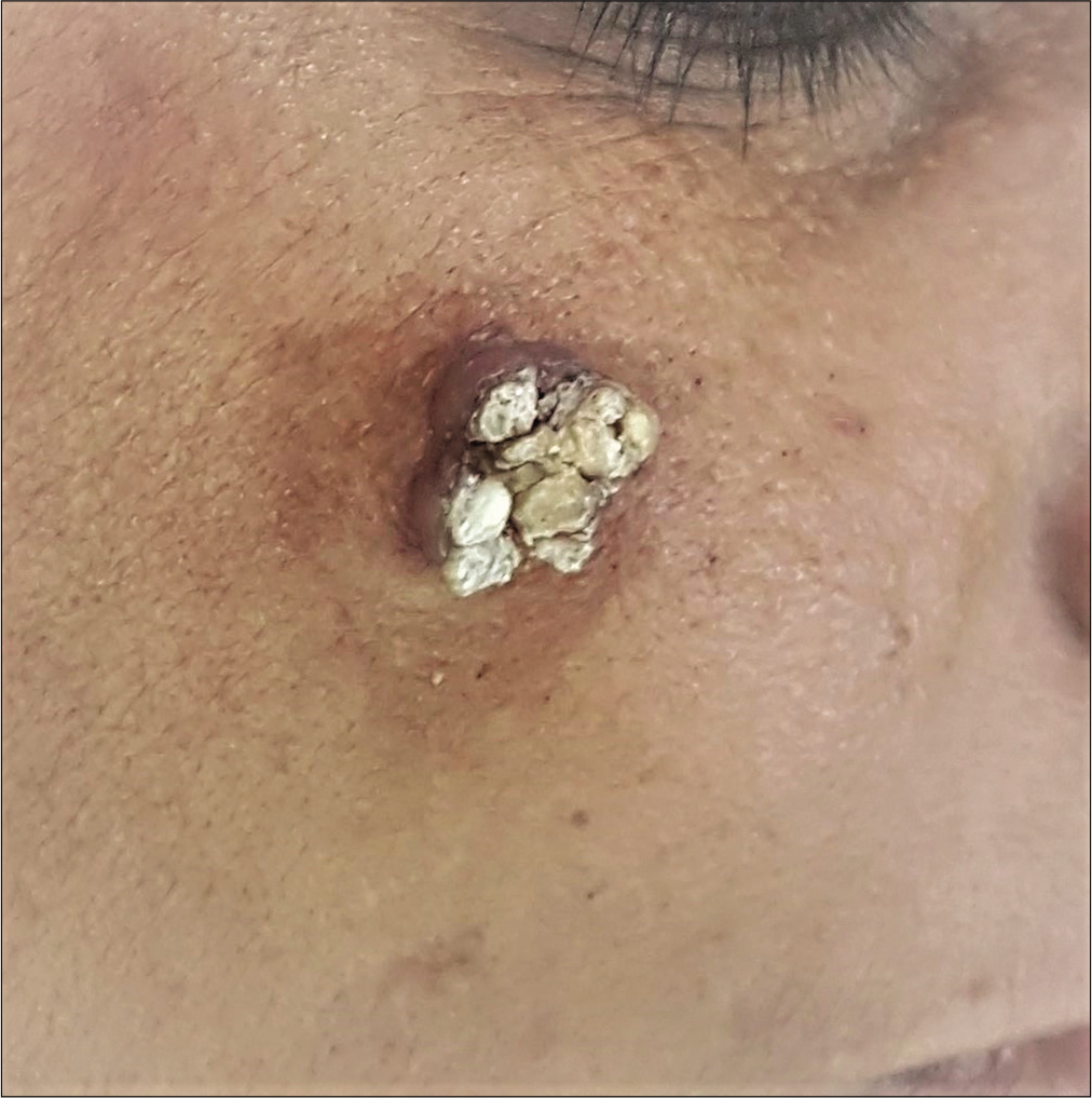
Cornified lesion of cutaneous horn on right cheek
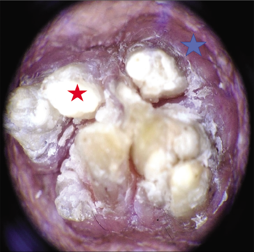
Figure 1:
Cornified lesion of cutaneous horn on right cheek
Multiple closely set off-white colored structures (red star) over a dull red colored background (blue star)
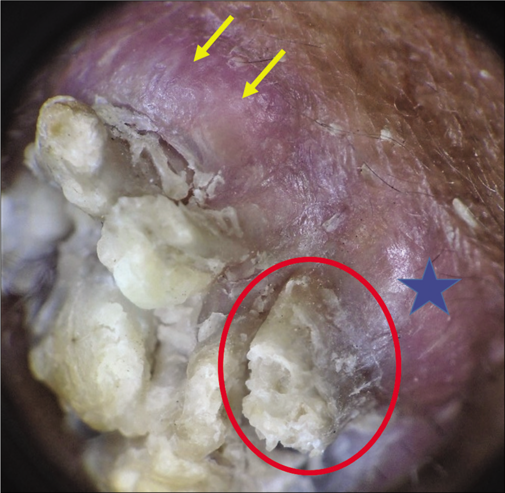
Figure 2a:
Multiple closely set off-white colored structures (red star) over a dull red colored background (blue star)
Side view of the horn (in red circle) showing the absence of structural horizontal contours (absent terrace phenomenon) and no specific vascular morphology and pattern in the background (blue star). Also, note the follicular scales (yellow arrow) (polarizing dermoscopy ×10)
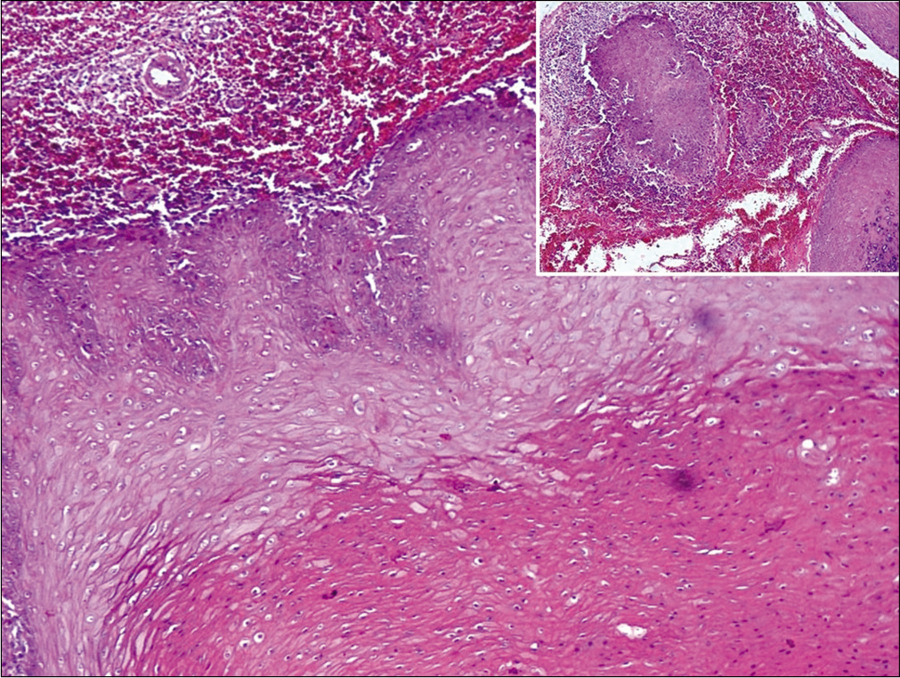
Figure 2b:
Side view of the horn (in red circle) showing the absence of structural horizontal contours (absent terrace phenomenon) and no specific vascular morphology and pattern in the background (blue star). Also, note the follicular scales (yellow arrow) (polarizing dermoscopy ×10)
The epidermis showing mounds of parakeratosis, acanthosis with koilocytosis and papillomatous rete ridges suggestive of invasive pathology (H&E, ×100). Inset shows dense lymphocytic infiltrate around the bulbous rete ridges
Figure 3:
The epidermis showing mounds of parakeratosis, acanthosis with koilocytosis and papillomatous rete ridges suggestive of invasive pathology (H&E, ×100). Inset shows dense lymphocytic infiltrate around the bulbous rete ridges

The present case unfolded the presence of verrucous carcinoma at the base of a cutaneous horn. Verrucous carcinoma (Ackerman’s tumor) is a well-differentiated warty variant of squamous cell carcinoma with low malignant potential. It typically involves the oral cavity, esophagus, larynx, genitalia and plantar surface of foot. Face is not a common site.4 Strong association with tobacco and betel nut chewing and HPV infection has been documented. There is a single study by Pyne et al. evaluating the dermoscopic features of cutaneous horn in 163 consecutive patients.5 The authors concluded that few dermoscopic features are significantly associated with underlying invasive SCC namely low height-to-base ratio, absence of terrace morphology and presence of base erythema and tenderness. They recorded lower height of the horn as compared to the diameter of the base in 67% cases of invasive squamous cell carcinoma. On the contrary, height of the horn was greater than base diameter in benign keratosis. All these features were seen in our patient. Terrace morphology is a feature presenting as structural horizontal contours on the side of the horn. On histopathology, it appears as ortho-hyperkeratosis with horizontal parallel lamellae of dead keratin. Base erythema is characterized by presence of red area within 5 mm of horn base that may be attributed to the increased perfusion. The close dermoscopic differentials include actinic keratosis, benign keratosis and SCC in situ. The differentiating dermoscopic features of cutaneous horn with underlying benign and malignant etiology have been tabulated in Table 1.

Table 1: Dermoscopic and clinical difference between benign and malignant horn
Feature Benign horn Malignant horn
Horn height: diameter of base Horn height more than the diameter of base Horn height less than base diameter
Terrace morphology Present Absent
Base erythema Less More
Pain on palpation Less More

Thus, cutaneous horns could be a harbinger of malignancy as seen in our case. Though histopathology remains the diagnostic gold standard, dermoscopy can offer a clue to the underlying malignant etiology and affect the decision of therapeutic and diagnostic surgical excision.

Declaration of patient consent

The authors certify that they have obtained all appropriate patient consent.

Financial support and sponsorship

Nil.

Conflicts of interest

There are no conflicts of interest.

References

  1. , , . Cutaneous horns: Are these lesions as innocent as they seem to be? World J Surg Oncol. 2004;2:18.
    [CrossRef] [PubMed] [Google Scholar]
  2. , , , . A histopathological study of 643 cutaneous horns. Br J Dermatol. 1991;124:449-52.
    [CrossRef] [PubMed] [Google Scholar]
  3. , , , , , . Cutaneous horn: A retrospective histopathological study of 222 cases. An Bras Dermatol. 2010;85:157-63.
    [CrossRef] [PubMed] [Google Scholar]
  4. , , . Verrucous carcinoma in a giant cutaneous horn: A case report and literature review. Case Rep Otolaryngol. 2020;2020:7134789.
    [CrossRef] [PubMed] [Google Scholar]
  5. , , . Cutaneous horns: Clues to invasive squamous cell carcinoma being present in the horn base. Dermatol Pract Concept. 2013;3:3-7.
    [CrossRef] [PubMed] [Google Scholar]

Fulltext Views
4,586

PDF downloads
1,758
View/Download PDF
Download Citations
BibTeX
RIS
Show Sections