Generic selectors
Exact matches only
Search in title
Search in content
Post Type Selectors
Search in posts
Search in pages
Filter by Categories
15th National Conference of the IAOMFP, Chennai, 2006
Abstract
Abstracts from current literature
Acne in India: Guidelines for management - IAA Consensus Document
Addendum
Announcement
Art & Psychiatry
Article
Articles
Association Activities
Association Notes
Award Article
Book Review
Brief Report
Case Analysis
Case Letter
Case Letters
Case Notes
Case Report
Case Reports
Clinical and Laboratory Investigations
Clinical Article
Clinical Studies
Clinical Study
Commentary
Conference Oration
Conference Summary
Continuing Medical Education
Correspondence
Corrigendum
Cosmetic Dermatology
Cosmetology
Current Best Evidence
Current Issue
Current View
Derma Quest
Dermato Surgery
Dermatopathology
Dermatosurgery Specials
Dispensing Pearl
Do you know?
Drug Dialogues
e-IJDVL
Editor Speaks
Editorial
Editorial Remarks
Editorial Report
Editorial Report - 2007
Editorial report for 2004-2005
Errata
Erratum
Focus
Fourth All India Conference Programme
From Our Book Shelf
From the Desk of Chief Editor
General
Get Set for Net
Get set for the net
Guest Article
Guest Editorial
History
How I Manage?
IADVL Announcement
IADVL Announcements
IJDVL Awards
IJDVL AWARDS 2015
IJDVL Awards 2018
IJDVL Awards 2019
IJDVL Awards 2020
IJDVL International Awards 2018
Images in Clinical Practice
Images in Dermatology
In Memorium
Inaugural Address
Index
Knowledge From World Contemporaries
Leprosy Section
Letter in Response to Previous Publication
Letter to Editor
Letter to the Editor
Letter to the Editor - Case Letter
Letter to the Editor - Letter in Response to Published Article
LETTER TO THE EDITOR - LETTERS IN RESPONSE TO PUBLISHED ARTICLES
Letter to the Editor - Observation Letter
Letter to the Editor - Study Letter
Letter to the Editor - Therapy Letter
Letter to the Editor: Articles in Response to Previously Published Articles
Letters in Response to Previous Publication
Letters to the Editor
Letters to the Editor - Letter in Response to Previously Published Articles
Letters to the Editor: Case Letters
Letters to the Editor: Letters in Response to Previously Published Articles
Media and news
Medicolegal Window
Messages
Miscellaneous Letter
Musings
Net Case
Net case report
Net Image
Net Images
Net Letter
Net Quiz
Net Study
New Preparations
News
News & Views
Obituary
Observation Letter
Observation Letters
Oration
Original Article
ORIGINAL CONTRIBUTION
Original Contributions
Pattern of Skin Diseases
Pearls
Pediatric Dermatology
Pediatric Rounds
Perspective
Presedential Address
Presidential Address
Presidents Remarks
Quiz
Recommendations
Regret
Report
Report of chief editor
Report of Hon : Treasurer IADVL
Report of Hon. General Secretary IADVL
Research Methdology
Research Methodology
Resident page
Resident's Page
Resident’s Page
Residents' Corner
Residents' Corner
Residents' Page
Retraction
Review
Review Article
Review Articles
Reviewers 2022
Revision Corner
Self Assessment Programme
SEMINAR
Seminar: Chronic Arsenicosis in India
Seminar: HIV Infection
Short Communication
Short Communications
Short Report
Snippets
Special Article
Specialty Interface
Studies
Study Letter
Study Letters
Supplement-Photoprotection
Supplement-Psoriasis
Symposium - Contact Dermatitis
Symposium - Lasers
Symposium - Pediatric Dermatoses
Symposium - Psoriasis
Symposium - Vesicobullous Disorders
SYMPOSIUM - VITILIGO
Symposium Aesthetic Surgery
Symposium Dermatopathology
Symposium-Hair Disorders
Symposium-Nails Part I
Symposium-Nails-Part II
Systematic Review and Meta-Analysis
Systematic Reviews and Meta-analyses
Systematic Reviews and Meta-analysis
Tables
Technology
Therapeutic Guideline-IADVL
Therapeutic Guidelines
Therapeutic Guidelines - IADVL
Therapeutics
Therapy
Therapy Letter
Therapy Letters
View Point
Viewpoint
What’s new in Dermatology
View/Download PDF

Translate this page into:

Letter to the Editor
2013:79:6;819-821
doi: 10.4103/0378-6323.120741
PMID: 24177620

Dermoscopic criteria for differentiating exogenous ochronosis from melasma

Niti Khunger, Rajat Kandhari
 Department of Dermatology and STD, V.M. Medical College and Safdarjang Hospital, New Delhi, India

Correspondence Address:
Niti Khunger
Department of Dermatology and STD, V.M. Medical College and Safdarjang Hospital, New Delhi - 110 029
India
How to cite this article:
Khunger N, Kandhari R. Dermoscopic criteria for differentiating exogenous ochronosis from melasma. Indian J Dermatol Venereol Leprol 2013;79:819-821
Copyright: (C)2013 Indian Journal of Dermatology, Venereology, and Leprology

Sir,

There has been a sudden rise in the number of reported cases of exogenous ochronosis (EO), [1],[2],[3] and it may not be as uncommon as previously thought. In the early stages, it is clinically difficult to distinguish EO from melasma. A worsening of pigmentation due to EO can lead to paradoxical increased application of skin lightening agents, further aggravating the condition. Thus, it is essential to distinguish early EO from melasma. The gold standard for diagnosis of EO is a skin biopsy. Dermoscopy may be an important tool to differentiate EO from melasma, and may assist in choosing the appropriate site for biopsy in suspected cases. Two cases of EO following the use of skin lightening agents for the treatment of melasma are reported and dermoscopic criteria for diagnosis are being put forward.

A 48-year-old Indian woman, Fitzpatricks skin type IV, presented with erythema, and gradual deterioration of her melasma. She was using sunscreens and skin lightening agents containing 2% hydroquinone in an unsupervised manner since 8 years. She noted worsening of her melasma since the past 3 years.

The second case was a 42-year-old, Indian woman, skin type IV diagnosed with melasma 14 years back. She was treated with modified Kligman′s formula containing 2% hydroquinone, 0.025% tretinoin, 1% hydrocortisone, sunscreens, and glycolic acid peels. She continued use of skin lightening agents unsupervised for 13 years, and presented with worsening of her melasma.

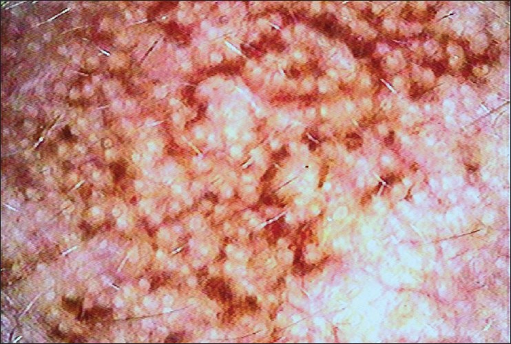
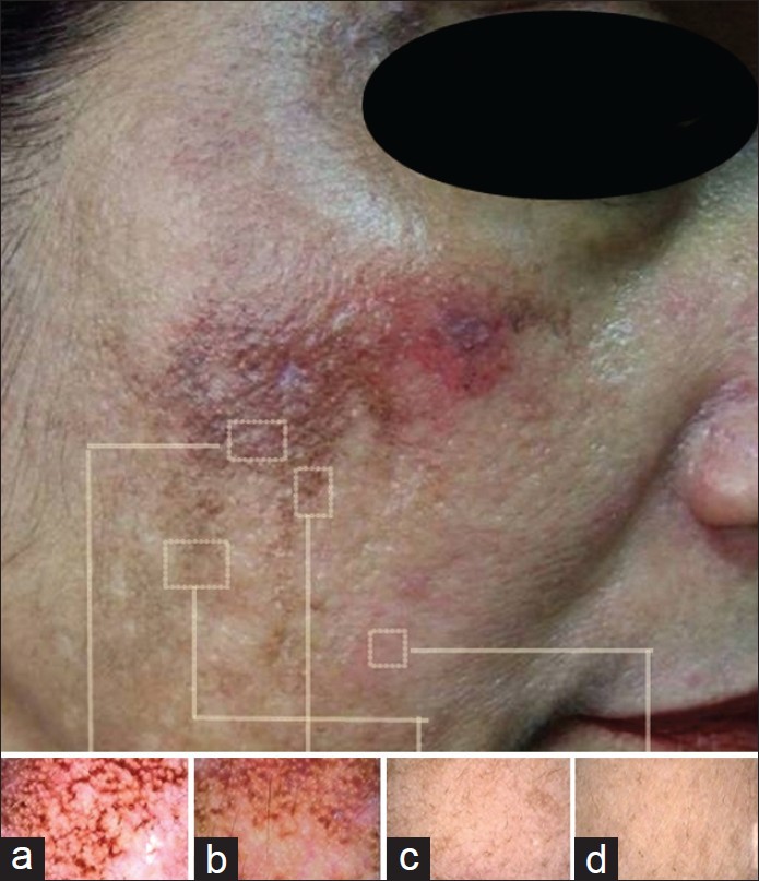
On clinical examination, both cases revealed grayish brown macules with interspersed "confetti like" hypo-pigmented macular areas on the malar region, a speckled pattern of pigmentation [Figure - 1]a-d and a coarse texture with pinpoint, dark brown papules, which were more appreciable on palpation [Figure - 1]d. In addition, case one, revealed erythema and fine telangiectasias on bilateral malar areas, whereas case two revealed mild atrophy of the malar regions and a mild bluish black hue of bilateral zygomatic regions. There was no clinical or laboratory evidence of alkaptonuria in both cases. 3 mm punch biopsies taken from the pinpoint papular lesions revealed characteristic short, stout, curvilinear, "banana-shaped," ochre-colored fibers of varying thickness in the papillary dermis [Figure - 1]f. Methylene blue staining showed dark blue staining of the ochronotic fibers [Figure - 1]e. There was evidence of solar elastosis in both cases. Both cases were clinical stage II as per Dogliotti staging. [4] Dermoscopic examination of both patients in areas with melasma without ochronosis revealed an accentuation of the normal pseudo-rete of the facial skin. In areas with ochronosis, greyish brown dark amorphous structures in the perifollicular region and some obliterating the follicular openings were observed. The pattern was curvilinear and "worm like" in some areas [Figure - 2]. There was a clear demarcation between melasma and exogenouss ochronosis on dermoscopy [Figure - 3]a-d.
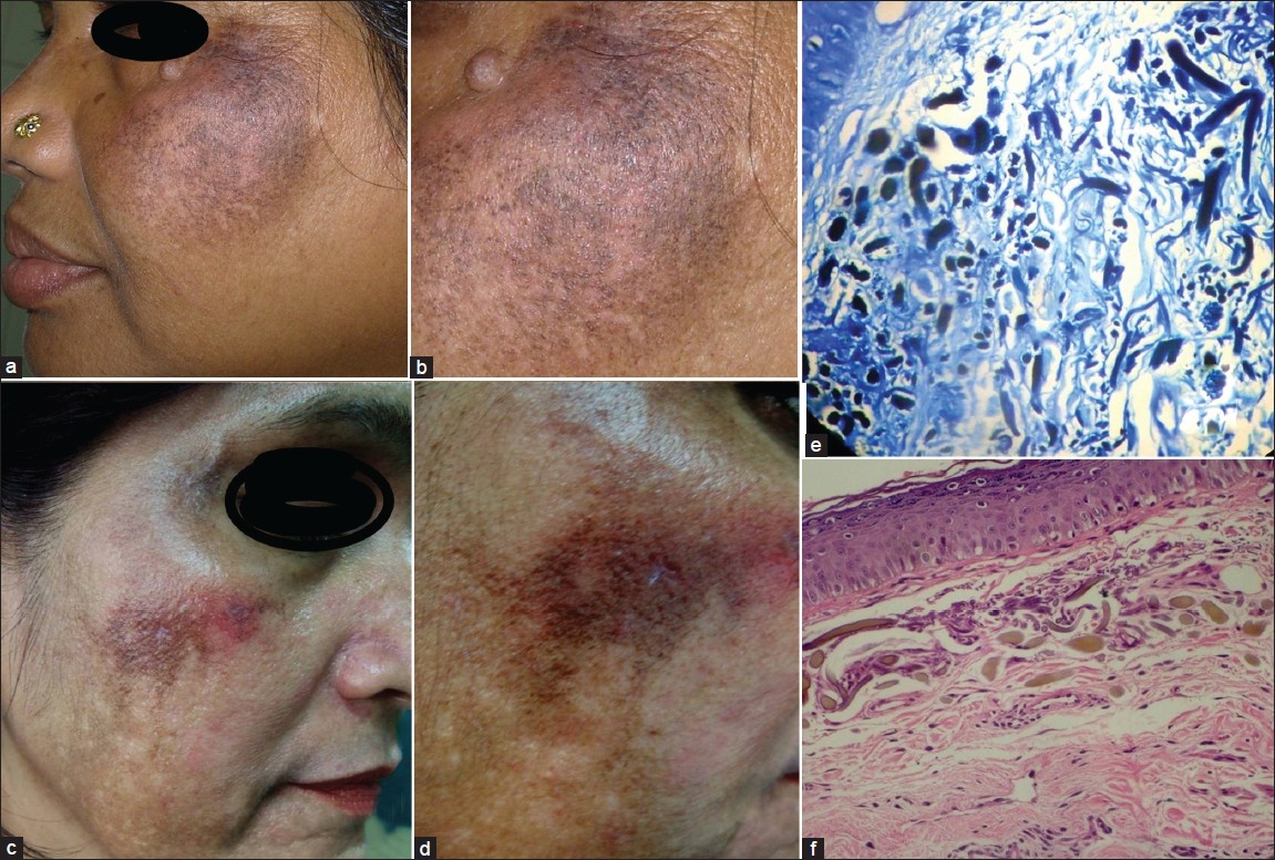
Figure 1: (a) Ochronosis in case 1 showing grayish brown macules with interspersed "confetti like" depigmented macular areas with erythema on the cheeks. (b) Close-up view showing speckled pattern of hyperpigmentation with a coarse texture and pinpoint, dark brown papules. (c) Bluish grey to brown macules and papules with interspersed "confetti like" depigmented macular areas. (d) Tiny pinpoint caviar like brown papules with telangiectasia. (e) Methylene blue staining showing ochronotic fi bers stained dark blue. (f) Characteristic short, stout, curvilinear, "banana-shaped," ochre-colored fi bers of varying thickness in the papillary and upper dermis
Figure 2: Dermoscopic features of ochronosis showing dark brown globules, elongated and curvilinear-worm like structures conjoined together in a reticulate pattern of ochronosis
Figure 3: (a) Ochronosis. (b) Sharp contrast between ochronosis in the upper half and melasma in the lower half. (c) Areas of melasma without ochronosis, showed accentuation of the normal pseudo-rete pattern with a diffuse brown pigmentation. (d) Normal skin

EO is clinically characterized by an asymptomatic hyperchromia of the skin, usually on the sun exposed areas of the face, back, and the extensor surfaces of the extremities. Tan et al. [5] recently reported variable clinical presentations of EO. Due to its varied presentation and striking similarity to melasma, especially in the early stages, clinicians require a high index of suspicion in order to make a diagnosis [Table - 1]. An early diagnosis is important as worsening of pigmentation may lead to application of increased concentration of hydroquinone rather than terminating it immediately.

Table 1: Clinical setting where dermoscopy should be used as a screening test

Charlín et al. [3] reported dermoscopic features of two patients with EO, wherein they observed blue-gray amorphous areas obliterating some follicular openings. Gil et al. [6] reported the dermoscopic features as irregular, brown-gray, globular, annular, and arciform structures. This was confirmed using a reflectance confocal microscope. Berman et al. [7] reported dark brown globules and globular-like structures on a diffuse brown background, in patients having EO, whereas those with melasma demonstrated a fine brown reticular pattern on a background of a faint light brown structure less area. Our dermoscopic findings were similar to previously reported findings. In addition, we observed a characteristic "worm-like" pattern. Thus, the dermoscopic features of EO are clearly distinct from melasma [Table - 2].

Table 2: Comparison of dermoscopic features of melasma and exogenous ochronosis

Hence, dermoscopy can be employed as a rapid screening test for EO. The clinical presence of coarse texture of the skin, fine telangiectasias and hyperchromia with "speckling" or "reticulation" [3] should alert the clinician to resort to a dermoscopic examination, particularly in patients who are reluctant to get a facial biopsy.

To conclude, dermoscopy can be used as a rapid, non-invasive tool to detect EO, and may be a useful guide in the selection for the appropriate site for biopsy in patients with pre-existing melasma.

References
1.
Tan SK. Exogenous ochronosis in ethnic Chinese Asians: A clinicopathological study, diagnosis and treatment. J Eur Acad Dermatol Venereol 2011;25:842-50.
[Google Scholar]
2.
Ribas J, Schettini AP, Cavalcante Mde S. Exogenous ochronosis hydroquinone induced: A report of four cases. An Bras Dermatol 2010;85:699-703.
[Google Scholar]
3.
Charlín R, Barcaui CB, Kac BK, Soares DB, Rabello-Fonseca R, Azulay-Abulafia L. Hydroquinone-induced exogenous ochronosis: A report of four cases and usefulness of dermoscopy. Int J Dermatol 2008;47:19-23.
[Google Scholar]
4.
Dogliotti M, Leibowitz M. Granulomatous ochronosis - A cosmetic-induced skin disorder in Blacks. S Afr Med J 1979;56:757-60.
[Google Scholar]
5.
Tan SK. Exogenous ochronosis - A diagnostic challenge. J Cosmet Dermatol 2010;9:313-7.
[Google Scholar]
6.
Gil I, Segura S, Martínez-Escala E, Lloreta J, Puig S, Vélez M, et al. Dermoscopic and reflectance confocal microscopic features of exogenous ochronosis. Arch Dermatol 2010;146:1021-5.
[Google Scholar]
7.
Berman B, Ricotti C, Vieira M, Amini S. Differentiation of exogenous ochronosis from melasma by dermoscopy. J Am Acad Dermatol. 2009;60(Suppl 1):AB2.
[Google Scholar]

Fulltext Views
14,298

PDF downloads
4,138
Show Sections