Translate this page into:
Eccrine angiomatous hamartoma with verrucous hemangioma-like features – an unusual combination
Corresponding author: Prof. Xian Jiang, Department of Dermatology, West China Hospital, Sichuan University, Sichuan, China. jennyxianj@163.com
-
Received: ,
Accepted: ,
How to cite this article: Liu L, Zhou L, Zhao Q, Wei D, Jiang X. Eccrine angiomatous hamartoma with verrucous hemangioma-like features – an unusual combination. Indian J Dermatol Venereol Leprol 2021;87:842-4.
Sir,
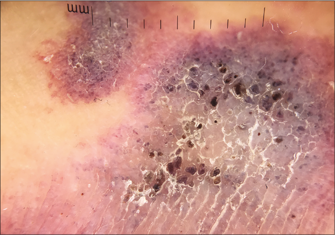
A 25-year-old male presented with a history of asymptomatic reddish-purple patches on the dorsal right hand since birth. On examination, there were several clustered, erythematousviolaceous and hyperkeratotic plaques on the dorsal right hand [Figure 1a]. Dermoscopy revealed a prominent blue-white background, with hyperkeratosis in the center of the lesions, surrounded by purple-brown round lacunae indicative of underlying dilated vessels [Figure 1b]. Doppler ultrasonography of the lesions detected dotted blood flow signals in the dermis and subcutis. Magnetic resonance imaging demonstrated mixed long T1 and T2 signals of lesions involving dermal and subcutaneous tissue without involving tendons and muscles, which was suggestive of a hemangioma.

- Multiple reddish-purple hyperkeratotic plaques on the dorsal right hand

- Dermoscopic examination: blue-white background, hyperkeratosis in the middle of the lesion, surrounded by purple-brown round lacunae
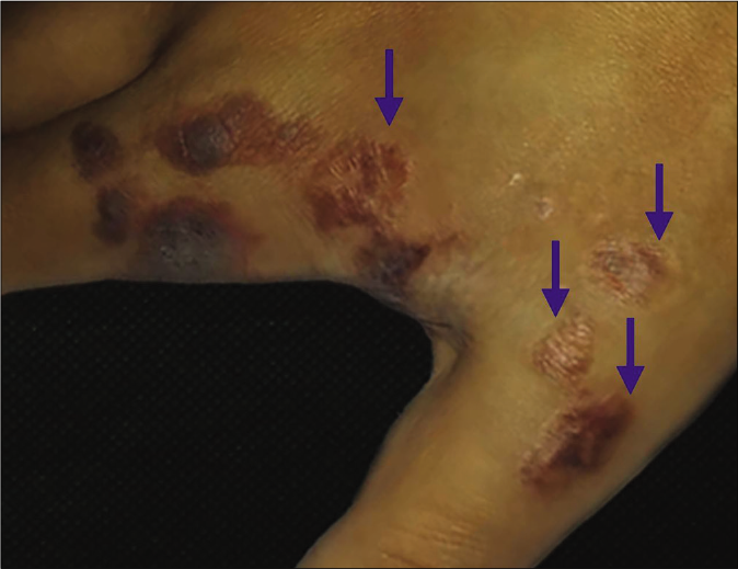
Skin biopsy showed hyperkeratosis, acanthosis and papillomatosis in the epidermis. The papillary dermis had numerous thin-walled, ectatic and irregular vessels [Figures 2a and 2b]. In the reticular dermis and subcutis, proliferation of eccrine glands associated with thin walled vessels was observed [Figure 2c]. On immunohistochemistry, the vascular endothelial cells were positive for CD31 [Figure 2d] and weakly positive for GLUT-1 [Figure 2e]. Positive attaining for αSMA was seen in the yoepithelial layer of secretory coils and vascular smooth muscle cells [Figure 2f]. We speculated that the diagnosis could be eccrine angiomatous hamartoma with verrucous hemangioma-like features. Intralesional Nd: YAG laser (energy 130 mJ, frequency 60 Hz) was utilized to treat the lesions which improved satisfactorily after two treatments [Figure 3].
![Histopathology examinations of lesions from dorsal aspect of the right hand. (a) Low-power view of biopsy (hematoxylin–eosin[HE],original magnification ×10). (b) High-power view showing hyperkeratosis, acanthosis and papillomatosis in epidermis,thin-walled, ectatic, vessels in papillary dermis (×100). (c) Proliferation of eccrine glands associated with thin-walled vessels in reticular dermis (×100). (d) Immunohistochemical staining showing that the vascular endothelial cells (green arrow) were positive for CD31 and eccrine glands (black arrow) were negative (×400). (e) Vessels were weakly positive for GLUT-1 (×400). (f) Myoepithelial layer of secretory coils (black arrow) and vessels (green arrow) were positive for αSMA (×400)](/content/126/2021/87/6/img/IJDVL-87-842-g003.png)
- Histopathology examinations of lesions from dorsal aspect of the right hand. (a) Low-power view of biopsy (hematoxylin–eosin[HE],original magnification ×10). (b) High-power view showing hyperkeratosis, acanthosis and papillomatosis in epidermis,thin-walled, ectatic, vessels in papillary dermis (×100). (c) Proliferation of eccrine glands associated with thin-walled vessels in reticular dermis (×100). (d) Immunohistochemical staining showing that the vascular endothelial cells (green arrow) were positive for CD31 and eccrine glands (black arrow) were negative (×400). (e) Vessels were weakly positive for GLUT-1 (×400). (f) Myoepithelial layer of secretory coils (black arrow) and vessels (green arrow) were positive for αSMA (×400)

- Apparent improvement of the plaques (blue arrow) after treatment with Nd:YAG laser
In a study of 26 cases of eccrine angiomatous hamartoma, seven demonstrated mild hyperkeratosis in the epidermis; another study involving 15 cases reported that one patient had verrucous changes in the epidermis and abundant capillaries in the papillary dermis.1,2 Wang et al. analyzed 74 cases of verrucous hemangioma and found hyperplasia of the eccrine glands around abnormal vessels in four cases. They opined that these were verrucous hemangiomas with features of eccrine angiomatous hamartoma.3 However, a literature search could find only two reported cases with the diagnosis of eccrine angiomatous hamartoma with verrucous hemangioma-like features. In Table 1 we have summarized the characteristics of eccrine angiomatous hamartoma and verrucous hemangioma so as to highlight their salient differences.].
| Features | Eccrine angiomatous hamartoma | Verrucous hemangioma |
|---|---|---|
| Onset | Congenital or later in childhood | Congenital or in early infancy |
| Location | Distal extremities | Distal extremities |
| Distribution | Mostly solitary papules | Grouped of plaques or nodules |
| Symptom | Pain, hyperhidrosis | Itch, oozing, bleeding |
| Dermoscopy | A spitzoid pattern or a popcorn pattern | Bluish-white hue (hyperkeratosis), reddish-blue or bluish lacunae |
| Histopathology | Eccrine sweat glands associated with thin-walled, aggregated vessels in the middle and lower dermis | Hyperkeratosis, acanthosis and papillomatosis in epidermis, vascular component in dermis and subcutaneous tissue |
| GLUT-1 expression | Negative | Mostly positive |
| Therapy | Surgery, laser, botulinum toxin | Surgery, laser, topical steroid with salicylic acid ointment |
Both eccrine angiomatous hamartoma and verrucous hemangioma present mostly at birth or childhood with lesions principally localized to the extremities. Eccrine angiomatous hamartoma is mostly isolated papules, while verrucous hemangioma is characterized as multiple and clustered plaques or nodules. The dermoscopic features of eccrine angiomatous hamartoma are spitzoid or popcorn patterns, whereas those of verrucous hemangioma are reddish-blue or bluish lacunae or dermis with a bluish-white hue. Clinically and dermoscopically the lesions seen in our patient were more suggestive of verrucous hemangioma. The symptoms of eccrine angiomatous hamartoma include pain and hyperhidrosis, whereas those of verrucous hemangioma are itch, oozing and bleeding. However, our patient did not exhibit typical symptoms of either entity.
Patterson et al. developed the histopathological criteria for eccrine angiomatous hamartoma including: (a) hyperplasia of normal or dilated eccrine glands; (b) intermixing of eccrine glands with abundant capillaries; (c) variable presence of apocrine, lymphatic or mucinous structures.4 Histology of verrucous hemangioma demonstrates hyperkeratosis, papillomatosis and acanthosis in the epidermis with numerous dilated vessels extending into the subcutaneous tissues. Our case showed histological characteristics of both eccrine angiomatous hamartoma and verrucous hemangioma.
GLUT-1 and WT-1 are markers that differentiate vascular neoplasms from vascular malformations. Trindade et al. reported that verrucous hemangioma was positive for GLUT-1 in 13 cases (100%), suggesting that verrucous hemangioma may be categorized as a form of vascular tumor.5 However, Wang et al. found that vessels in verrucous hemangioma were positive for GLUT-1 in 49 cases (66%), focally positive for Prox1 in 69 (93%) cases, while negative for WT-1 in 60 cases (81%).3 They proposed that verrucous hemangioma is a vascular malformation and an incomplete lymphatic immunophenotype. The reason why for GLUT-1 is positive in verrucous hemangioma is not clear, but the staining is less intense than in infantile hemangiomas.
In conclusion, the mixed histological features comprising both eccrine gland proliferation as well as vascular proliferation and weak GLUT-1 positivity support the diagnosis of eccrine angiomatous hamartoma with verrucous hemangioma-like features in our case. To the best of our knowledge, this is the first reported case of eccrine angiomatous hamartoma with verrucous hemangioma-like features, to be characterized in such great detail using a combination of histopathology, immunohistochemistry and dermoscopy.
Declaration of patient consent
The authors certify that they have obtained all appropriate patient consent.
Financial support and sponsorship
National Natural Science Foundation of China (Grant No. 81872535 and 82003373).
Conflicts of interest
There are no conflicts of interest.
References
- Eccrine angiomatous hamartoma: A clinicopathological study of 26 cases. Dermatology. 2015;231:63-9.
- [CrossRef] [Google Scholar]
- Eccrine angiomatous hamartoma: A retrospective study of 15 cases. Chang Gung Med J. 2012;35:167-77.
- [CrossRef] [Google Scholar]
- Verrucous hemangioma: A clinicopathological and immunohistochemical analysis of 74 cases. J Cutan Pathol. 2014;41:823-30.
- [CrossRef] [Google Scholar]
- Eccrine angiomatous hamartoma: A clinicopathologic review of 18 cases. Am J Dermatopathol. 2016;38:413-7.
- [CrossRef] [Google Scholar]
- An immunohistochemical study of verrucous hemangiomas. J Cutan Pathol. 2013;40:472-6.
- [CrossRef] [Google Scholar]


