Translate this page into:
Enhancing syphilis diagnosis through innovative adaptation of wet mount microscopy
Corresponding author: Dr. Senkadhir Vendhan, Department of Dermatology, Babasaheb Ambedkar Memorial Hospital, Byculla, Mumbai, Maharashtra, India. vendhan100@gmail.com
-
Received: ,
Accepted: ,
How to cite this article: Vendhan S, Vasudevan B, Bala K, Neema S. Enhancing syphilis diagnosis through innovative adaptation of wet mount microscopy. Indian J Dermatol Venereol Leprol. 2024;90:844-5. doi: 10.25259/IJDVL_374_2024
Problem
The diagnosis of syphilis relies on direct tests like dark ground illumination (DGI) and indirect serological assays.1 Dark ground microscopy is known for its reliability in detecting the syphilis-causing Treponema pallidum due to its unique morphology and motility but is inaccessible in many centres.2 This poses a significant challenge for timely and accurate diagnosis of syphilis, especially in resource-limited settings. Some authors have recommended using dark-field mode in advanced light microscopes and employing plastic sheets or coins to block the light source.3 However, the advantage of employing this method in identifying Treponema species has not been mentioned.
Solution
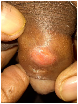
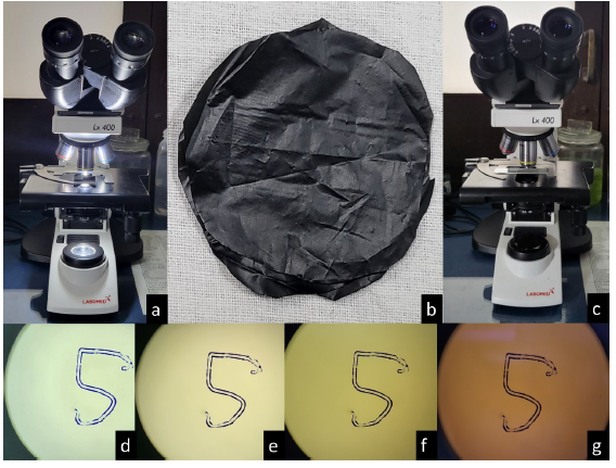
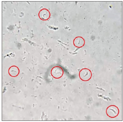
A novel approach has been developed based on insights from three cases of primary syphilis that involved adapting routine microscopy with specific modifications to detect Treponema pallidum. Serum samples from the moist lesions of syphilis are smeared on the glass slides and secured with a coverslip [Figure 1]. A modified microscope setup is then utilised, incorporating a thin, black plastic cover between the light source and the glass slide to reduce background light interference. We used three plastic covers one over the other [Figure 2]. Additionally, a dark room is created by blocking all external light sources, and the intensity of the microscope’s light source is minimised to facilitate the observation of characteristic spirochete movements [Video 1]. To minimise potential disruptions from air bubble movements, ventilation systems such as fans are turned off during observation. Notably, the distinctive corkscrew movement of the spirochete aids in their identification [Figure 3 and Video 2]. Practical recommendations include emphasis on the importance of sample collection and prompt microscopic examination with a minimal light source to prevent to enhance diagnostic accuracy and sensitivity. This innovative adaptation offers a promising alternative for syphilis diagnosis in settings where traditional dark ground microscopy is not available. A limitation of this innovation is that it cannot match the contrast achieved with a dark-field microscope.

- Clinical image showing solitary ulcer over the glans.

- (a-c) show the steps for converting a normal microscope into a modified dark ground microscope, (d) displays the microscopy field without a black cover, (e) with a single layer, (f) with a double layer, and (g) with a three-layer cover.

- Wet mount microscopy (400x) shows spiral structures (highlighted in red circles) indicative of spirochetes.
Declaration of patient consent
The authors certify that they have obtained all appropriate patient consent.
Financial support and sponsorship
Nil.
Conflicts of interest
There are no conflicts of interest.
Use of artificial intelligence (AI)-assisted technology for manuscript preparation
The authors confirm that there was no use of artificial intelligence (AI)-assisted technology for assisting in the writing or editing of the manuscript and no images were manipulated using AI.
References
- The laboratory diagnosis of syphilis. Can J Infect Dis Med Microbiol. 2005;16:45-51.
- [CrossRef] [PubMed] [PubMed Central] [Google Scholar]
- A new method in demodex imaging: Shining demodex in the dark field mode of the new generation digital light microscope. Skin Res Technol. 2023;29:13361.
- [CrossRef] [PubMed] [PubMed Central] [Google Scholar]


