Translate this page into:
Epidermal nevus in blaschkoid distribution treated with dual-wavelength copper vapor laser
Corresponding author: Dr. Igor V. Ponomarev, 53, Leninskiy Prospect, Moscow, 119991, Russian Federation. iponom@okb.lpi.troitsk.ru
-
Received: ,
Accepted: ,
How to cite this article: Ponomarev IV, Topchiy SB, Kluchareva SV, Shakina LD. Epidermal nevus in blaschkoid distribution treated with dual-wavelength copper vapor laser. Indian J Dermatol Venereol Leprol 2021;87:718-20.
Sir,
Epidermal nevi develop due to benign overgrowth of cells either involving keratinocytes only (non-organoid type) or in combination with cells of hair follicles, sebaceous glands or sweat glands (organoid type). Epidermal Blaschko line-distributed nevi may be represented by linear verrucous inflammatory nevus, sebaceous and non-organoid nevus. Linear verrucous inflammatory nevus appears as red, eczematous-like area.
Epidermal nevus usually occurs on the trunk and limbs and is uncommon on the face or scalp.1 Blaschkoid epidermal nevi are associated with post-zygotic mutations.2
Facial epidermal blaschkoid nevus without both inflammation and itching, clinically, may be consistent with the diagnosis of nevus sebaceous. On clinical grounds, such case should be presented as an epidermal nevus in blaschkoid distribution.
Facial blaschkoid nevus gives rise to poor cosmetic appearance and makes patients seek the aid of dermatologists to remove it. Surgical excision of facial blaschkoid nevus may be ineffective and associated with recurrences and scar formation. The ablative lasers (Er: YAG, CO2) were used for the treatment of small- or medium-sized nevus sebaceous with acceptable results. The use of CO2 laser for the treatment of nevus sebaceous has shown fair results, but is associated with hyperpigmentation, mild atrophic scarring and significant downtime as side effects.3,4 The treatment of nevus sebaceous should include total elimination of acanthotic cells as well as sebocytes and remodeling of the adjacent vascular bed of the involved area. The dual-wavelength copper vapor laser wavelengths (511 and 578 nm) are highly absorbed by melanin, lipids and blood chromophores and seem to be optimal for the removal of nevus sebaceous, targeting all relevant chromophores in the lesional acanthotic epidermal cells, sebocytes and blood vessels.5 This is a report of the use of copper vapor laser to successfully treat facial epidermal blaschkoid nevi which has not been reported hitherto.
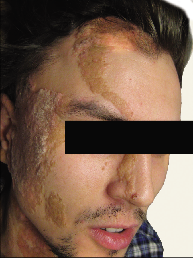
A 24-year-old man with Fitzpatrick skin phototype II presented with a yellowish plaques, sized 10 × 20 cm, along Blaschko’s lines on the right cheek, forehead, nose, neck [Figure 1a] and back of the trunk. Located on the face, this patch showed neither any inflammation nor itching. Based on clinical signs (as the photomicrograph is not available), this case may be considered as epidermal nevus in blaschkoid distribution.

- Epidermal nevus in blaschkoid distribution over the face in a male patient before copper vapor laser treatment
The patient had no associated growth or developmental abnormalities such as ocular, neurological or bone anomalies.
The informed consent was obtained after the patient was counseled regarding the risks and benefits of laser treatment. At the request of the patient, laser treatment was carried out only on the face.
The facial and scalp lesions were treated with copper vapor laser (Yakhroma-Med, Lebedev Physical Institute of the Russian Academy of Sciences). The settings were as follows: average power of 0.8–1.0 W, a ratio at green (511 nm) and yellow (578 nm) wavelengths of 3:2, the exposure time was 0.2 s and the diameter of the light spot on the skin was 1 mm. The treatment endpoint was the treated area acquiring a grayish tint. The treatment was performed without anesthesia. After the procedure, the skin was treated with 0.05% solution of chlorhexidine gluconate and Bepanthen cream twice a day.
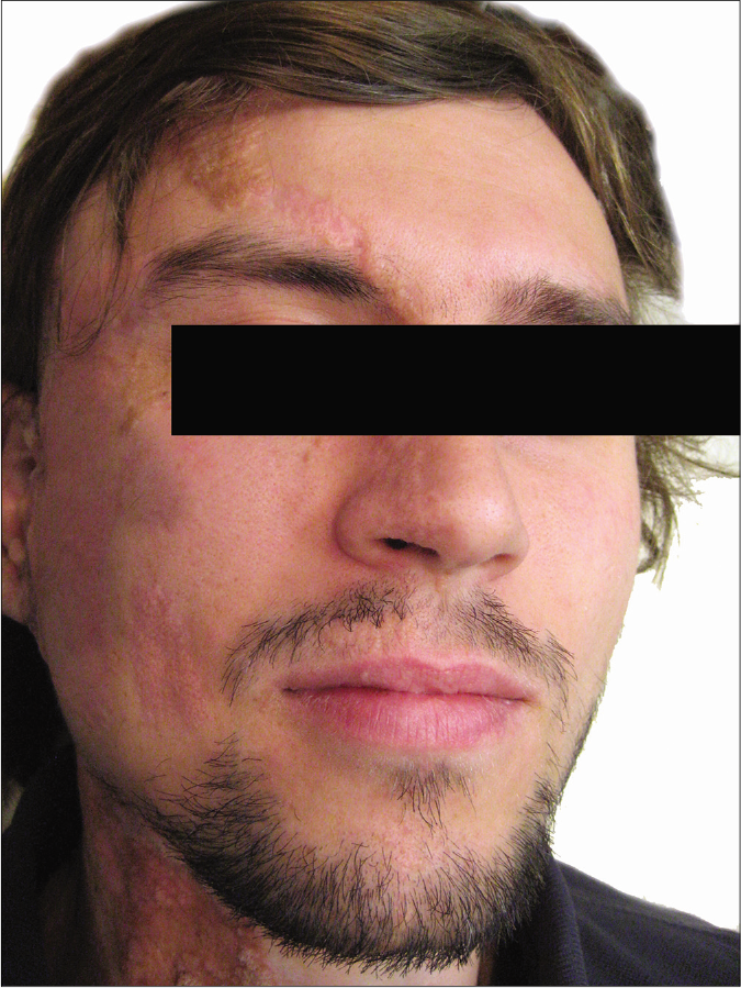
The laser procedure was not accompanied by any bleeding or erythema. The irradiated skin healed with exfoliation after ten days with complete restoration of epidermis, without any pigmentary changes. The lesions were treated with copper vapor laser six times at an interval of two months. Following six sessions of dual-wavelength laser radiation, the facial nevus sebaceous compartment was completely cleared [Figure 1b]. No recurrences were observed up to 24 months after the final laser treatment.

- Fair resolution of the plaque eight months after six copper vapor laser treatments
In the laser management of the epidermal blaschkoid nevus, the target chromophores include melanin in acanthotic cells, lipids in sebocytes as well as oxyhemoglobin and hemoglobin in erythrocytes of the microvascular bed of the papillary dermis.6 The high simultaneous absorption of dual-wavelength copper vapor laser radiation at 511 nm and 578 nm by all the target nevus sebaceous chromophores provided the sound removal of nevus sebaceous due to the appropriate heating of melanin in acanthotic cells, sufficient sebum discharge and photodestruction of the involved dilated microvascular bed.7 Copper vapor laser at 578 nm, mostly absorbed by oxyhemoglobin and deoxyhemoglobin, provides appropriate photocoagulation followed by the remodeling of the vascular bed, likely preventing both relapses and malignant change in facial blaschkoid nevus.7 The limited penetration depth of copper vapor laser in the dermis due to the high absorption of radiation by melanin, sebum lipids, oxyhemoglobin and hemoglobin determines the main advantage of copper vapor laser in comparison with other laser systems. Copper vapor laser neither passes into the deep dermis nor overheats dermal stem cells, essential for the appropriate skin healing after the laser exposure.6 Copper vapor laser can be useful in the treatment of facial epidermal nevi distributed along Blaschko’s line without inflammatory signs.
Additional prospective studies with more patients and longer follow-up are warranted to determine the treatment efficacy.
Declaration of patient consent
The authors certify that they have obtained all appropriate patient consent.
Financial support and sponsorship
Nil.
Conflicts of interest
There are no conflicts of interest.
References
- Linear lesions in dermatology. Indian J Dermatol Venereol Leprol. 2011;77:722-6.
- [CrossRef] [Google Scholar]
- Blaschko lines and other patterns of cutaneous mosaicism. Clin Dermatol. 2011;29:205-25.
- [CrossRef] [Google Scholar]
- Evaluation of carbon dioxide laser in the treatment of epidermal nevi. J Cutan Aesthet Surg. 2016;9:183-7.
- [CrossRef] [Google Scholar]
- Nevus sebaceous: Response to erbium YAG laser ablation. Indian J Plast Surg. 2005;38:48-50.
- [CrossRef] [Google Scholar]
- Optical properties of biological tissues: A review. Phys Med Biol. 2013;58:R37-61.
- [CrossRef] [Google Scholar]
- Rhinophyma treatment by copper vapor laser with the computerized scanner. J Lasers Med Sci. 2019;10:153-6.
- [CrossRef] [Google Scholar]
- Treatment of basal cell cancer with a pulsed copper vapor laser: A case series. J Lasers Med Sci. 2019;10:350-4.
- [CrossRef] [Google Scholar]


