Translate this page into:
Epithelioid sarcoma of the extremities
2 Department of Dermatology, Kafkas University, Kars, Turkey
3 Department of Pathology, Cerrahpasa Faculty of Medicine, University of Istanbul, Istanbul, Turkey
4 Departments of Plastic and Reconstructive Surgery, Dr. Lutfi Kirdar Kartal Training and Research Hospital, Istanbul, Turkey
5 Departments of Plastic and Reconstructive Surgery, Faculty of Medicine, Bezmialem Vakif University, Istanbul, Turkey
Correspondence Address:
Fatma Akpinar
Maltepe �niversitesi Tıp Fak�ltesi Dermatoloji Anabilim Dalı Atat�rk Cd. �am Sk. No:3 34843 Maltepe/ İstanbul
Turkey
| How to cite this article: Akpinar F, Dervis E, Demirkesen C, Akpinar AC, Ergun SS. Epithelioid sarcoma of the extremities. Indian J Dermatol Venereol Leprol 2014;80:168-170 |
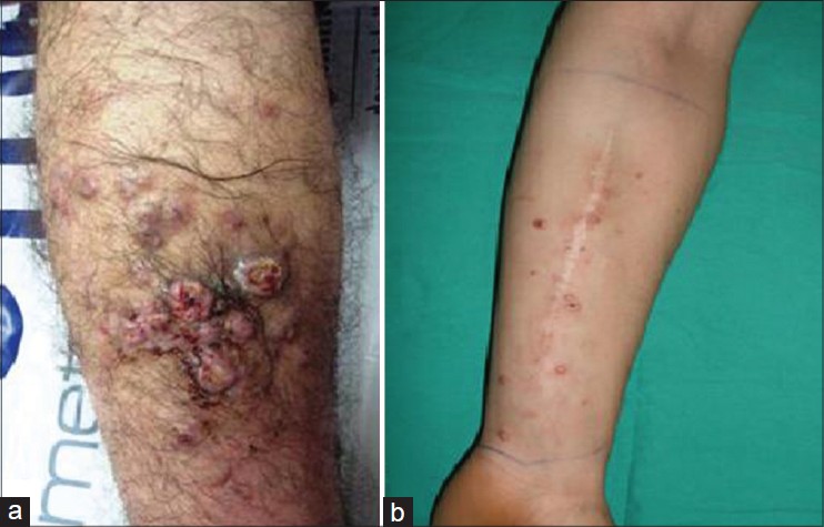
Case 1: A 27-year-old man presented with an asymptomatic nodule on his right lower leg of one year duration, which he believed could be a malignant tumour. He was treated with topical medication; however the nodule progressively enlarged and new nodules appeared in the surrounding skin over a few months. On examination,there were multiple, 1 × 2 cm ulcerating nodules on the mid-anterior aspect of the right lower leg, around which there were satellite nodules along with right inguinal lymphadenopathy [Figure - 1]a.
 |
| Figure 1: (a) Case 1: Ulcerating nodules on the mid-anterior aspect of the right lower leg. (b) Case 2: Multiple, small nodules on the anterior aspect of the left forearm |
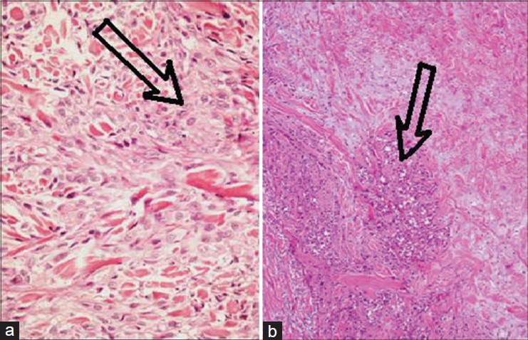
Histopathogical examination of the punch biopsy specimen taken from the nodule revealed collections of epithelioid cells around areas of necrosis in the dermis [Figure - 2]a. Immunohistochemistry was reactive for pancytokeratin and nonreactive for CD20, CD3, CD34, CD31, CD68, CD1a [Figure - 3]a, suggestive of epithelioid sarcoma. Serum CA125 level was elevated. Thoraco-abdominal computerised tomography showed no evidence of metastasis. Magnetic resonance imaging revealed a 12 × 2.5 × 3 cm irregular mass in the antero-medial aspect of the right lower leg, invading the soleus muscle and fascia.
 |
| Figure 2: (a) Case 1: Focal areas of necrosis in the dermis accompanied by diffusely arranged epithelioid cells (on both sides of the base of the arrow) and focal collection of cells around necrotic areas, suggestive of granuloma-like structures (around the head of the arrow) (H and E, ×100). (b) Case 2: Pseudogranulomatous arrangement of epithelioid and fusiform cells (in front of the head of the arrow) (H and E, ×40) |
 |
| Figure 3: (a) Case 1: The majority of dermal infi ltrating cells were positive for pancytokeratin. (b) Case 2: Immunohistochemical expression of cytokeratin. (c) Case 2: Immunohistochemical expression of vimentin |
After a multi-disciplinary consultation involving the specialities of orthopedics, plastic surgery, dermatology and oncology, transfemoral amputation of the right leg along with inguinal lymph node dissection was performed. Histopathological examination of the excised specimen revealed bone invasion and metastasis in 3 of 15 lymph nodes examined. Six cycles of chemotherapy with a regimen comprising ifosfamide, cisplatin, vincristine and doxorubicin was administered. The patient was followed-up for a year during which time, there was no regional or distant metastasis detected.
Case 2: A 30-year-old male patient presented with a swelling over his left forearm, which slowly progressed in size over 2 years. On clinical examination, there was an immobile nodule of size 3 × 4 cm on the anterior aspect of the left forearm. Incisional biopsy of the nodule was performed and histopathological examination (HPE) revealed acanthosis and spindle-shaped and epithelioid cells surrounding a necrotic zone in the reticular dermis. έmmunohistochemical analysis revealed that the cells were positive for vimentin, CD34, epithelial membrane antigen and cytokeratin, consistent with epithelioid sarcoma.
Magnetic resonance imaging did not show invasion of fascia. Computerised tomography of the forearm showed no evidence of metastasis. Complete excision of the nodule with safe surgical margins was done. Histopathology of the excised specimen confirmed the diagnosis of epithelioid sarcoma. One year later, he presented with multiple nodules at the same site [Figure - 1]b. Punch biopsy of one of the nodules revealed features suggestive of local recurrence of the sarcoma. No evidence of local invasion was found on imaging studies; however, ultrasonography demonstrated enlargement of the axillary lymph nodes.
After multidisciplinary review, above-elbow amputation and excision of the axillary lymph nodes was undertaken. Histopathology demonstrated pseudo-granulomatous arrangement of epithelioid and fusiform cells [Figure - 2]b. Immunohistochemical studies revealed co-expression of cytokeratin [Figure - 3]b and vimentin [Figure - 3]c. The patient is on regular follow up in the department of oncology.
Epithelioid sarcoma is a slow-growing malignancy with a high frequency of recurrence and metastasis. The mean duration of symptoms reported by Chase and Enzinger in 1985 is 29 months (range: 1 week to 25 years). [1] It is often misdiagnosed at the initial visit. [2] It is mostly seen in young adults aged between 20 and 40 years old. [3] It usually involves the dermis, subcutis, or deeper soft tissues in the distal extremities. [2] Often, the superficial lesions ulcerate leading to a mistaken diagnosis of a poorly healing traumatic wound or wart. The demographic characteristics and clinical features of our patients were consistent with previous reports in the literature. Our cases were initially misdignosed as nonspecific ulcers and treated with antibiotics but no improvement was observed. Unlike most other sarcomas, epithelioid sarcoma has a tendency to spread locally by way of lymphatics or along fascial planes and may give rise to multiple local nodules. [4] It has a tendency to develop metastases to lung, bone, brain and other locations. [5]
The paucity of symptoms, a benign appearance in early-stage imaging and indistinctive histopathology in some cases makes the diagnosis challenging. In our patients, the tumours progressed asymptomatically for months before detection. Biopsy is the diagnostic modality of choice. The tumor consists of epithelioid cells well blended with fusiform cells. The most common pattern seen is a "pseudogranulomatous" proliferation of cells around a necrotic zone. It exhibits immunohistochemical reactivity for keratins and epithelial membrane antigen, vimentin and CD34 (in more than half of the cases). [5]
One of our patients, in addition, had a high level of CA 125. Serum CA 125 level could be a useful marker for diagnosis and for monitoring the clinical course.
Histopathological differential diagnoses for epithelioid sarcoma include both benign and malignant conditions such as granuloma annulare, necrobiosis lipoidica, fibrous histiocytoma, nodular fasciitis, melanoma, clear-cell sarcoma of the tendon and aponeurosis, metastatic squamous cell carcinoma, synovial sarcoma, epithelioid hemangioendothelioma and malignant extra-renal rhabdoid tumors of the soft tissue. [5] The multiple differential diagnoses frequently cause a delay in diagnosis.
Epithelioid sarcoma should be considered in the differential diagnosis of chronic nodules, especially on the extremities of young men.
| 1. |
Burgos AM, Chávez JG, Sánchez JL, Sánchez NP. Epithelioid sarcoma: A diagnostic and surgical challenge. Dermatol Surg 2009;35:687-91.
[Google Scholar]
|
| 2. |
Chase DR, Enzinger FM. Epithelioid sarcoma. Diagnosis, prognostic indicators, and treatment. Am J Surg Pathol 1985;9: 241-63.
[Google Scholar]
|
| 3. |
de Visscher SA, van Ginkel RJ, Wobbes T, Veth RP, Ten Heuvel SE, Suurmeijer AJ, et al. Epithelioid sarcoma: Still an only surgically curable disease. Cancer 2006;107:606-12.
[Google Scholar]
|
| 4. |
Pai KK, Pai SB, Sripathi H, Pranab, Rao P. Epithelioid sarcoma: A diagnostic challenge. Indian J Dermatol Venereol Leprol 2006;72:446-8.
[Google Scholar]
|
| 5. |
Armah HB, Parwani AV. Epithelioid sarcoma. Arch Pathol Lab Med 2009;133:814-9.
[Google Scholar]
|
Fulltext Views
2,917
PDF downloads
1,561


