Generic selectors
Exact matches only
Search in title
Search in content
Post Type Selectors
Search in posts
Search in pages
Filter by Categories
15th National Conference of the IAOMFP, Chennai, 2006
Abstract
Abstracts from current literature
Acne in India: Guidelines for management - IAA Consensus Document
Addendum
Announcement
Art & Psychiatry
Article
Articles
Association Activities
Association Notes
Award Article
Book Review
Brief Report
Case Analysis
Case Letter
Case Letters
Case Notes
Case Report
Case Reports
Clinical and Laboratory Investigations
Clinical Article
Clinical Studies
Clinical Study
Commentary
Conference Oration
Conference Summary
Continuing Medical Education
Correspondence
Corrigendum
Cosmetic Dermatology
Cosmetology
Current Best Evidence
Current Issue
Current View
Derma Quest
Dermato Surgery
Dermatopathology
Dermatosurgery Specials
Dispensing Pearl
Do you know?
Drug Dialogues
e-IJDVL
Editor Speaks
Editorial
Editorial Remarks
Editorial Report
Editorial Report - 2007
Editorial report for 2004-2005
Errata
Erratum
Focus
Fourth All India Conference Programme
From Our Book Shelf
From the Desk of Chief Editor
General
Get Set for Net
Get set for the net
Guest Article
Guest Editorial
History
How I Manage?
IADVL Announcement
IADVL Announcements
IJDVL Awards
IJDVL AWARDS 2015
IJDVL Awards 2018
IJDVL Awards 2019
IJDVL Awards 2020
IJDVL International Awards 2018
Images in Clinical Practice
Images in Dermatology
In Memorium
Inaugural Address
Index
Knowledge From World Contemporaries
Leprosy Section
Letter in Response to Previous Publication
Letter to Editor
Letter to the Editor
Letter to the Editor - Case Letter
Letter to the Editor - Letter in Response to Published Article
LETTER TO THE EDITOR - LETTERS IN RESPONSE TO PUBLISHED ARTICLES
Letter to the Editor - Observation Letter
Letter to the Editor - Study Letter
Letter to the Editor - Therapy Letter
Letter to the Editor: Articles in Response to Previously Published Articles
Letters in Response to Previous Publication
Letters to the Editor
Letters to the Editor - Letter in Response to Previously Published Articles
Letters to the Editor: Case Letters
Letters to the Editor: Letters in Response to Previously Published Articles
Media and news
Medicolegal Window
Messages
Miscellaneous Letter
Musings
Net Case
Net case report
Net Image
Net Images
Net Letter
Net Quiz
Net Study
New Preparations
News
News & Views
Obituary
Observation Letter
Observation Letters
Oration
Original Article
ORIGINAL CONTRIBUTION
Original Contributions
Pattern of Skin Diseases
Pearls
Pediatric Dermatology
Pediatric Rounds
Perspective
Presedential Address
Presidential Address
Presidents Remarks
Quiz
Recommendations
Regret
Report
Report of chief editor
Report of Hon : Treasurer IADVL
Report of Hon. General Secretary IADVL
Research Methdology
Research Methodology
Resident page
Resident's Page
Resident’s Page
Residents' Corner
Residents' Corner
Residents' Page
Retraction
Review
Review Article
Review Articles
Reviewers 2022
Revision Corner
Self Assessment Programme
SEMINAR
Seminar: Chronic Arsenicosis in India
Seminar: HIV Infection
Short Communication
Short Communications
Short Report
Snippets
Special Article
Specialty Interface
Studies
Study Letter
Study Letters
Supplement-Photoprotection
Supplement-Psoriasis
Symposium - Contact Dermatitis
Symposium - Lasers
Symposium - Pediatric Dermatoses
Symposium - Psoriasis
Symposium - Vesicobullous Disorders
SYMPOSIUM - VITILIGO
Symposium Aesthetic Surgery
Symposium Dermatopathology
Symposium-Hair Disorders
Symposium-Nails Part I
Symposium-Nails-Part II
Systematic Review and Meta-Analysis
Systematic Reviews and Meta-analyses
Systematic Reviews and Meta-analysis
Tables
Technology
Therapeutic Guideline-IADVL
Therapeutic Guidelines
Therapeutic Guidelines - IADVL
Therapeutics
Therapy
Therapy Letter
Therapy Letters
View Point
Viewpoint
What’s new in Dermatology
View/Download PDF

Translate this page into:

Letter to the Editor
2012:78:3;376-377
doi: 10.4103/0378-6323.95467

Extensive blue nevus of the pharynx

Wen-Sen Lai1 , Chiang-Hung Hsu1 , Jih-Chin Lee2 , Yueng-Hsiang Chu2
1 Department of Otolaryngology-Head and Neck Surgery, Tri Service General Hospital, National Defense Medical Center, Taipei; Taichung Armed forces General Hospital, Taichung, Taiwan, Republic of China,
2 Department of Otolaryngology-Head and Neck Surgery, Tri Service General Hospital, National Defense Medical Center, Taipei, Taiwan, Republic of China,

Correspondence Address:
Yueng-Hsiang Chu
Department of Otolaryngology-Head and Neck Surgery, Tri Service General Hospital, National Defense Medical Center, 325, Sec. 2, Cheng-Kung Rd., Taipei 114, Taiwan, Republic of China

How to cite this article:
Lai WS, Hsu CH, Lee JC, Chu YH. Extensive blue nevus of the pharynx. Indian J Dermatol Venereol Leprol 2012;78:376-377
Copyright: (C)2012 Indian Journal of Dermatology, Venereology, and Leprology

Sir,

The blue nevus is a benign, acquired melanocytic lesion that typically presents as an asymptomatic, slate-blue or blue-black, smooth-surfaced macule or papule, measuring less than 6 mm in diameter usually. [1] This lesion occurs mostly on the skin and rarely in the oral mucosa. [2] It is the most common type of nevus occurring in the oral cavity after intramucosal nevi and accounts for 19% to 36% of oral nevi. [3] Intraoral blue nevus was first described in 1959. Intraoral blue nevi occur most frequently on the hard palate, with the buccal mucosa being the second most common site. [2] We describe a case of blue nevus of the pharyngeal mucosa. To the best of our knowledge, this is the first report on blue nevi occurring in this unusual site.

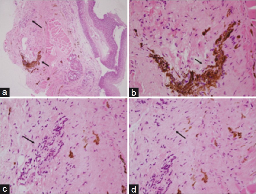
A widespread blue-black plaque was detected in the oropharyngeal mucosa of a 58-year-old woman during a routine health examination. The patient was aware of the asymptomatic lesion for more than 20 years. She did not have pigmented lesions on any other part of the body and there was no history of trauma, infection, or addiction to tobacco. There were no pigmented lesions on the mucosal surfaces of the eyes, genitalia, or digestive tract. The results of the remaining clinical examinations were not remarkable. Endoscopy using a 0-degree rigid nasopharyngoscope and a 70-degree rigid laryngoscope revealed an irregularly shaped pigmented lesion in the oropharynx extending superiorly to the nasopharynx and inferiorly to the hypopharynx [Figure - 1]. The overlying mucosa was smooth, with no swelling or bulge. A tissue specimen was obtained from the posterior oropharyngeal wall by a punch biopsy. Histopathological examination of the obtained specimen showed infiltration of non-nested dermal dark spindle melanocytes associated with fibrosis, which are consistent with blue nevus [Figure - 2].
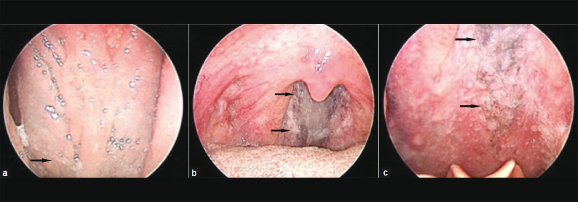
Figure 1: The scope revealed an irregularly shaped pigmented area (black arrows) extending superiorly to the level of the nasopharynx (a) and inferiorly to the level of the hypopharynx (c), with the main part lying in the oropharynx (b)
Figure 2: H and E stained section of the lesion at ×40 magnification (a) and ×400 magnification (b, c and d). A benign proliferation of spindle melanocytes (a and c, long arrows), with prominent melanin pigmentation (a and b, short arrows). The spindle melanocytes were infiltrated with fibroblasts (d, arrow)

Nevocytic nevi are protean lesions composed of several cell types arranged in a variety of histologic patterns. Dermal melanocytosis encompasses a spectrum of lesions including the following: (1) Mongolian spot, (2) nevus of Ota, (3) acquired nevus-of-Ota-like macules, (4) acquired dermal melanocytosis of the face and extremities, (5) nevus of Ito, (6) dermal melanocyte hamartoma, and (7) blue nevus. [4] The blue nevus is distinguished from the others on the basis of its histological presentation of a distorted dermis characterized by a greater concentration of dendritic melanocytes together with an increased fibrous tissue and melanophage. However, the lesions listed above are histologically similar to some extent. Thus, the diagnosis of dermal melanocytosis should be confirmed by taking into consideration both the histological picture and the clinical features of the lesion such as size, surface, and location.

Intraoral melanocytic nevi are uncommon and mostly occur on the palate or gingiva, although any mucosal site may be involved. The most common melanocytic nevi occurring in the oral cavity are of the intramucosal type (63%), followed by blue nevi (19%), compound nevi (9%), junction nevi (5%), and combined nevi (4%). [5]

Blue nevi generally occur on the skin of the hands, feet, and buttocks; however, in rare instances, they may occur in mucous membranes, including the oral mucosa. In most cases, blue nevi occurring in the oral mucosa are less than 10 mm in diameter and located on the hard palate. The common blue nevus, which is the most frequent subtype seen in the oral cavity, is characterized by the intramucosal proliferation of elongated, bipolar, spindle-shaped melanocytes that are often grouped in short fascicles arranged parallel to the overlying epithelium. Histologically, the pigmented spindle melanocytes can also be found in malignant melanoma and Spitz nevus.

In the differential diagnosis of the lesion, a melanoma arising form the pharyngeal mucosa is first considered, the other diseases include pseudomelanoma (recurrent melanocytic nevus), nicotine keratoses, drug induced pigmentation, oral melanoacanthoma, pigmentation induced by dental amalgams, nevus of o0 ta, and Mongolian spots, etc. The monomorphic and bland appearance of the proliferating cells and the absence of both mitotic activity and an infiltrating growth pattern ruled out melanocytic malignancy. In addition, the bluish pigmentation lesion in the pharyngeal mucosa should be distinguished from different types of hemangiomas, such as blue rubber-bleb nevus. In this case, the patient showed neither bluish vascular nevi of the skin nor hemangiomas of the gastrointestinal tract.

In conclusion, for both cutaneous and noncutaneous blue nevi, the preferred treatment is surgical excision, which generally results in a cure. However, a complete excision was not possible in this case because of the extent and location of the lesion. Melanotic lesions are rare in the pharyngeal mucosa. Diagnostic ENT endoscopes and ultrasonogrpahy in this case were very illustrative in demonstrating the extensive blue cell nevus of the pharynx, which was never hitherto formerly delineated.

References
1.
Scofield HH. The blue (Jadassohn-Tieche) nevus: A previously unreported intraoral lesion. J Oral Surg Anesth Hosp Dent Serv 1959;17:4-14.
[Google Scholar]
2.
Fistarol SK, Itin PH. Plaque-type blue nevus of the oral cavity. Dermatology 2005;211:224-33.
[Google Scholar]
3.
Buchner A, Hansen LS. Pigmented nevi of the oral mucosa: A clinicopathologic study of 36 new cases and review of 155 cases from the literature. Part II: Analysis of 191 cases. Oral Surg Oral Med Oral Pathol 1987;63:676-82.
[Google Scholar]
4.
Scolyer RA, Thompson JF, Stretch JR, Sharma R, McCarthy SW. Pathology of melanocytic lesions: New, controversial, and clinically important issues. J Surg Oncol 2004;86:200-11.
[Google Scholar]
5.
Buchner A, Hansen LS. Pigmented nevi of the oral mucosa: A clinicopathologic study of 32 new cases and review of 75 cases from the literature. Part I. A clinicopathologic study of 32 new cases. Oral Surg Oral Med Oral Pathol 1979;48:131-42.
[Google Scholar]

Fulltext Views
2,171

PDF downloads
1,255
Show Sections