Translate this page into:
Extralesional vitiligo over knee following halo nevus with poliosis on the scalp
Correspondence Address:
Uday Khopkar
Department of Dermatology, Seth GS Medical College and KEM Hospital, Parel, Mumbai-400 012
India
| How to cite this article: Gutte R, Khopkar U. Extralesional vitiligo over knee following halo nevus with poliosis on the scalp. Indian J Dermatol Venereol Leprol 2011;77:522-525 |
Sir,
A localized patch of gray or white hair is called poliosis. It can be associated with a variety of diseases, drugs, and lesions ranging from benign to malignant. [1] Halo nevus (HN) also known as Sutton′s nevus is a pigmented lesion usually melanocytic surrounded by a halo of depigmenattion. [2] HN over scalp with poliosis is rarely reported. [3]
Here, we present a case of HN with poliosis over the scalp preceding the development of vitiligo at a distant site.
Our patient, a 22-year-old male, presented with a complaint of circumscribed area of depigmentation and white hairs gradually increasing since the last 3 years over the occipital region of scalp around a raised black lesion which was present since birth. He had developed a patch of depigmentation on the left knee since the last 2 years.
On examination, a sharply demarcated halo of depigmentation with poliosis of overlying hair was seen around a variably pigmented plaque [Figure - 1]. On the knee there was a patch of circumscribed depigmentation of around 2 cm diameter with leucotrichia over and around the patch with accentuation on Wood′s lamp examination [Figure - 2].
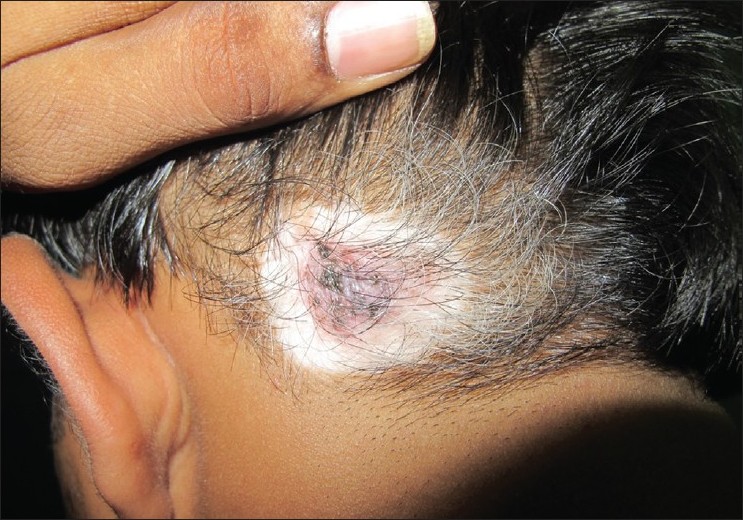 |
| Figure 1: A sharply circumscribed area of depigmentation with poliosis and pigmented plaque in center with areas of regression |
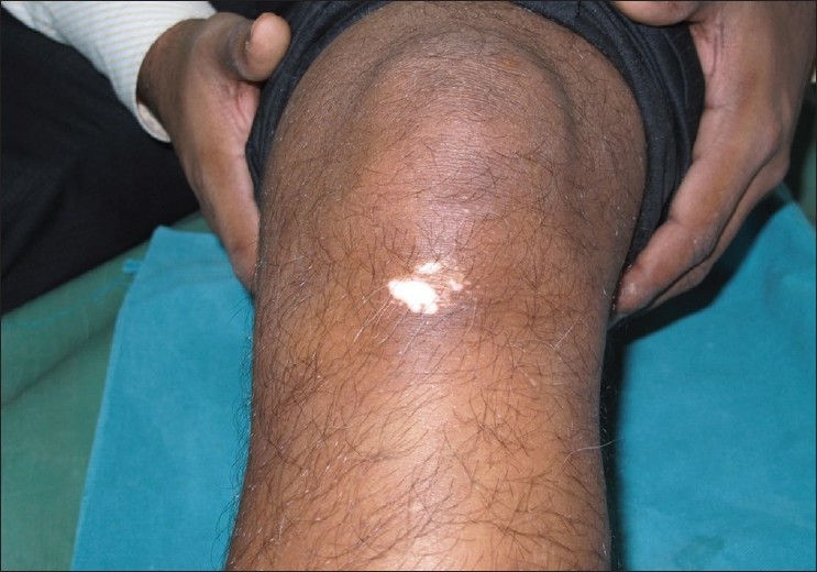 |
| Figure 2: A patch of vitiligo with leucotrichia on the left knee |
Biopsy done from the depigmented halo showed decreased number of melanocytes with sparse superficial perivascular lymphocytic infiltrate [Figure - 3] and that from the pigmented center showed multiple nests of melanocytes both within the epidermis and dermis along with moderately dense lymphocytic infiltrate around the nests suggestive of HN. Infiltrate was also extending to hair follicles, and follicular melanocytes appeared to be decreased [Figure - 4] and [Figure - 5]. The patient was referred to plastic surgery for excision of the lesion due to cosmetic concern of the patient.
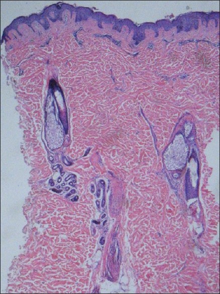 |
| Figure 3: Biopsy from depigmented halo showing decreased pigmentation and melanocytes in the epidermis with perivascular lymphocytic infiltrate (H and E, ×100) |
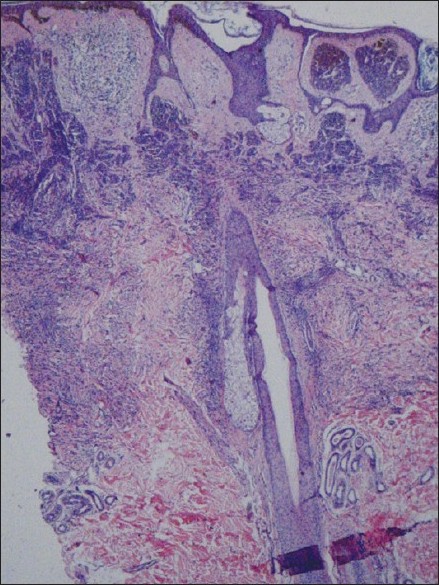 |
| Figure 4: Biopsy from pigmented lesion showing compound melanocytic nevus with diffuse lymphocytic infiltrate. (H and E, ×100) |
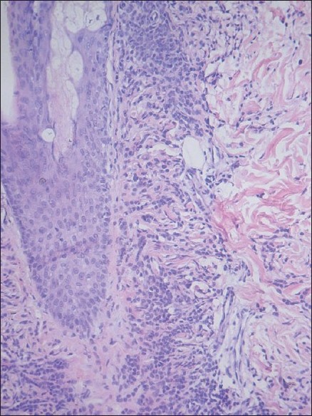 |
| Figure 5: Lymphocytic infiltrate extending to hair follicle and mixed within melanocytic nests (H and E, ×200) |
HN was described by Sutton as leucoderma acquisitum centrifugum and is also known as Sutton′s nevus. It commonly occurs over the back and may affect adults or children. Although it usually occurs as a single lesion in up to 50% cases, multiple lesions may be seen. [2],[4]
Histopathologically, it shows junctional, intradermal, or compound melanocytic nevus with variable atypia and a surrounding inflammatory infiltrate of T-lymphocytes. There are also some cases of noninflammatory halo nevi in which there is no inflammatory infiltrate histologically and such nevi do not involute. [4],[5] In addition, there are some cases in which the nevus shows inflammation histologically but no halo around it clinically, the so-called HN phenomenon or HN without a halo. [5]
HN is rarely reported to occur over scalp with poliosis. [3] Poliosis was the initial symptom that drew the patient′s attention to the underlying depigmentation in our case. Poliosis is known to occur with a giant congenital melanocytic nevus, congenital intradermal nevus, blue nevus, neurofibroma, nevus depigmentosus, and HN. [2],[3] Thus, poliosis may be a pointer to an underlying condition and its presence warrants thorough examination for underlying cause. Poliosis of eyelashes as an unusual and presenting sign of a HN has been reported. [6]
Halo nevi leading to the development of extralesional vitiligo have been reported. Development of vitiligo may precede, follow, or may be simultaneous with the development of halo around the nevus. Hofmann et al.[7] reported simultaneous development of segmental vitiligo and a halo surrounding a CMN.
Occurrence of vitiligo after the occurrence of HN in our patient lends credence to the theory that a HN if not excised in time may be a harbinger of vitiligo. Excision of HN to prevent development of vitiligo should be done as early as possible before the vitiligo has started. Association between these two conditions is more likely to be interrelated than coincidental, and immunological factors seem to be crucial for development of both halo phenomenon and vitiligo. The presence of cytotoxic CD8-positive T cells is probably responsible for regression of nevus due to direct cytotoxic damage, and emigration of these cells may be responsible for vitiligo lesions at sites distant from HN. [7]
In summary, appearance of depigmented halo and poliosis around a congenital melanocytic nevus and extralesional vitiligo with leucotrichia in the same individual as in our case suggests common immunological mechanism of these conditions, probably associated with melanocyte-specific cytotoxic T cells that can attack not only nevus cells but also epidermal and follicular melanocytes.
| 1. |
Young LC, Van Dyke GS, Lipton S, Binder SW. Poliosis overlying a nevus with blue nevus features. Dermatol Online J 2008;14:20.
[Google Scholar]
|
| 2. |
Patrizi A, Neri I, Sabattini E, Rizzoli L, Misciali C. Unusual inflammatory and hyperkeratotic halo nevus in children. Br J Dermatol 2005;152:357-60.
[Google Scholar]
|
| 3. |
Fellman AC, Mehregen AH. Letter: Halo nevi of scalp with poliosis. Arch Dermatol 1976;112:559.
[Google Scholar]
|
| 4. |
Mooney MA, Barr RJ, Buxton MG. Halo nevus or halo phenomenon? A study of 142 cases. J Cutan Pathol 1995;22:342-8.
[Google Scholar]
|
| 5. |
Lever WF, Schaumburg-Lever G. On benign melanocytic tumors and malignant melanoma. In: Lever WF, Schaumburg-Lever G, editors. Histopathology of the skin. 7 th ed. New York: Lippincott; 1990. p. 756-805.
[Google Scholar]
|
| 6. |
Kay KM, Kim JH, Lee TS. Poliosis of eyelashes as an unusual sign of halo nevus. Korean J Ophthalmol 2010;24:237-9.
[Google Scholar]
|
| 7. |
Hofmann UB, Brocker EB, Hamm H. Simultaneous onset of segmental vitiligo and a halo surrounding a congenital melanocytic nevus. Acta Derm Venereol 2009;89:402-6
[Google Scholar]
|
Fulltext Views
5,374
PDF downloads
1,774


