Translate this page into:
Genital squamous cell carcinoma presenting with multicentric ulcers: An unusual manifestation of skin malignancy
2 Department of Dermatology, Rize University, Rize, Turkey
3 Department of Pathology, Rize University, Rize, Turkey
Correspondence Address:
Pelin �st�ner
Dermatology Clinic, Rize State Hospital, Eminettin Caddesi, Hastaneler Kavsagi 53100, Rize
Turkey
| How to cite this article: �st�ner P, Dilek N, G��er H. Genital squamous cell carcinoma presenting with multicentric ulcers: An unusual manifestation of skin malignancy. Indian J Dermatol Venereol Leprol 2012;78:195-197 |
Sir,
Advanced genital tumors are very rare. Herein, we report a case of genital squamous cell carcinoma (SCC) accompanying a long-standing, well-healed scar tissue mimicking a venereal disease.
A 61-year-old white man presented with painful, exudative, firm pubic ulcers of 6 months duration. He described an asymptomatic wide scar-like lesion that developed in his genital area after improvement of an abscess 6 months ago and also informed that wide nonhealing ulcers developed on his right inguinal region 5 months ago. His medical history was unremarkable. He was a married man who retired from a textile company, and he denied any history of suspicious sexual behavior or encounters.
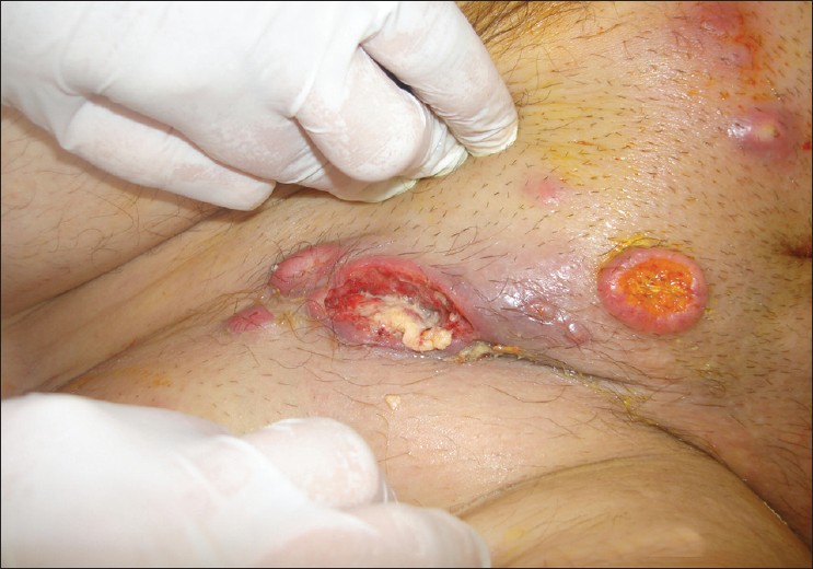
Dermatological examination revealed multiple painful, deep, punched-out ulcers (0.5-2 cm) with a central yellowish exudation and fungating, ulcero-proliferative growths with everted edges fixed to the underlying tissues [Figure - 1]. Another rapidly growing and hemorrhagic, wide and perforating ulcer, 5 cm × 6 cm in size, like a bubo, with a foul-smelling discharge, was also noted on his right inguinal region [Figure - 2]. The inguinal lymph nodes on the left were enlarged, hard and mobile.
 |
| Figure 1: Multiple painful, deep, punched-out ulcers (0.5-2 cm) with a yellowish exudation on the center and fungating, ulcero-proliferative growths with everted edges fixed to the underlying tissues |
 |
| Figure 2: Wide and perforating ulcer in 5 cm × 6 cm-like buboes, bleeding on touch with a foul-smelling discharge on his right inguinal region |
Routine investigations including hemogram were within normal limits. Anti-HBc, anti-HCV, anti-HIV and VDRL were negative. The bacterial, fungal, tuberculosis cultures, the Gram stain and the acid-resistant stains of the ulcerated lesion were all negative. The complement fixation tests, direct fluorescent antibody tests and the polymerase chain reaction (PCR) test from the ulcerated lesion for Chlamydia trachomatis and human pappiloma virus (HPV) were also negative. After informed consent, incisional biopsy was obtained from the ulcer edge. Histopathological examination revealed a poorly differentiated SCC. An infiltration of squamous cells, atypical, anaplastic, rounded cells with a focus of necrosis and dyskeratosis, were seen in the dermis (H and E, ×200) [Figure - 3]. Thorax tomography revealed bilateral multinodular metastatic lung lesions and abdominal tomography showed a soft tissue mass consisting of multiple centrally hypodense, necrotic lymphadenopathies in 0.5-2 cm diameter in the right parailiac region. Wide excision and split-thickness skin grafting of the pubic area and block resection of the ilio-inguinal lymph nodes were carried out. The histopathological examination of the resected specimen was suggestive of poorly differentiated SCC. Eight out of eight inguinal lymph nodes removed showed metastases bilaterally. Adjuvant radiotherapy of 50 Gy in fractionated doses and a combination chemotherapy protocol including methotrexate, bleomycin and cisplatinum were given for four courses with an uneventful recovery.
 |
| Figure 3: A dermal infiltration of islands consisting of squamous cells, atypical, anaplastic, rounded cells with focus of necrosis and dyskeratosis (H and E, × 200) |
In the literature, there are only a few publications of genital SCC accompanied by a chronic scar tissue. [1],[2] A 65-year-old man was reported to be diagnosed with SCC 2 years after a Fournier′s gangrene on the right hemiscrotum. [1] Kotwal et al. reported a 63-year-old Indian male who presented with a painless, firm lump over the penoscrotal junction diagnosed as SCC where he had a scar from a previous operation for infertility 30 years before. [3] Three cases of vulvar SCC that developed in episiotomy scars were also reported. [2]
Multicentric and multifocal anogenital SCC are rare, with the presence of HPV 16 DNA in Southern blot hybridization in most of them. [4] The multicentricity of the lesions in the anogenital SCC might be due to the multifocal invasion of the HPV lesions after autoinoculation on the anogenital region. [5] However, the absence of HPV in the lesional biopsy by PCR was another point for discussion in this case.
The development of genital cancer in this case could probably be explained by the synergistic effect of chronic irritation secondary to a prior fungal infection or poor hygiene rather than industrial exposure.
We wish to draw attention to the possibility of the malignancy in the differential diagnosis of long-standing, unresponsive, recurrent ulcerated lesions on the genital region. A chronic tender nodule or an ulcer on the genital area could easily be mistaken for an inflammatory lesion or a venereal disease. Therefore, low threshold for histopathological examination is mandatory to make an early diagnosis. Venereal diseases like lymphogranuloma venereum (LGV) or chancroid may be entertained in the differential diagnosis of this case, given the fact that tender bubo-like inguinal ulcers are present. In addition, cutaneous metastasis from another malignancy should also be considered due to the multiplicity of the lesions.
| 1. |
Chintamani, Shankar M, Singhal V, Singh JP, Bansal A, Saxena S. Squamous cell carcinoma developing in the scar of Fournier's gangrene case report. BMC Cancer 2004;4:16.
[Google Scholar]
|
| 2. |
Van Dam PA, Irvine L, Lowe DG, Fisher C, Barton DP, Shepherd JH. Carcinoma in episiotomy scars. Gynecol Oncol 1992;44:96-100.
[Google Scholar]
|
| 3. |
Kotwal S, Madaan S, Prescott S, Chilka S, Whelan P. Unusual squamous cell carcinoma of the scrotum arising from a well healed, innocuous scar of an infertility procedure: A case report. Ann R Coll Surg Engl 2007;89:17-9.
[Google Scholar]
|
| 4. |
Ikenberg H, Göppinger A, Hilgarth M, Pfisterer J, Müller U, Hillemanns HG, et al. Human papillomavirus, type 16, DNA in multicentric anogenital neoplasia associated with idiopathic panmyelopathy. A case report. J Reprod Med 1993;38:820-2.
[Google Scholar]
|
| 5. |
Beckmann AM, Acker R, Christiansen AE, Sherman KJ. Human papillomavirus infection in women with multicentric squamous cell neoplasia. Am J Obstet Gynecol 1991;165:1431-7.
[Google Scholar]
|
Fulltext Views
6,721
PDF downloads
1,707


