Translate this page into:
Giant congenital Becker's nevus overlying a plexiform neurofibroma: Merely a coincidence or more than it?
2 Department of Pathology, All India Institute of Medical Sciences, New Delhi, India
Correspondence Address:
Sujay Khandpur
Department of Dermatology and Venereology, All India Institute of Medical Sciences, New Delhi
India
| How to cite this article: Singh S, Khandpur S, Rai M, Ali F. Giant congenital Becker's nevus overlying a plexiform neurofibroma: Merely a coincidence or more than it?. Indian J Dermatol Venereol Leprol 2018;84:97-100 |
Sir,
Neurofibromas are commonly associated with various pigmentary skin lesions. Becker's nevus overlying a neurofibroma has been rarely described in the literature. Here, we report a rare case of giant congenital Becker's nevus overlying a plexiform neurofibroma in a young man without any manifestations of neurofibromatosis type I.
A 22-year-old man presented with an asymptomatic, non-progressive, large hyperpigmented patch over the back since birth. At the age of 15 years, he started developing coarse terminal hair on this hyperpigmented patch associated with multiple asymptomatic papular lesions. Since 2 years, he also appreciated an insidiously increasing, asymptomatic, soft swelling on mid-back under the hyperpigmented patch. Examination showed large brown, macule measuring 54 × 34 cms with splashed geographic margin, involving almost the entire back, with increased coarse terminal hair over it. On the mid-back, there was an ill-defined, 30 × 18 cms, elevated soft plaque with a 'bag of worm' feeling on palpation. Hairs were relatively sparse on the plaque. Multiple acneiform papules were also seen on the patch [Figure - 1]. Other mucocutaneous sites and systemic examination were within normal limits. A biopsy from the soft plaque showed a pan-dermal spindle cell proliferation with slender, thin, wavy, tapering nuclei. There were multiple mast cells interspersed between spindle cells [Figure 2a] and [Figure 2b]. Special staining with S100 showed strong positivity in the mid and deep dermis [Figure - 3]. Overall, histopathology was suggestive of neurofibroma. Biopsy from the hyperpigmented macule showed elongated and squared rete ridges with basal layer hyperpigmentation, suggestive of Becker's nevus [Figure - 4]. On clinicopathologic correlation, diagnosis of giant Becker's nevus overlying a plexiform neurofibroma was made.
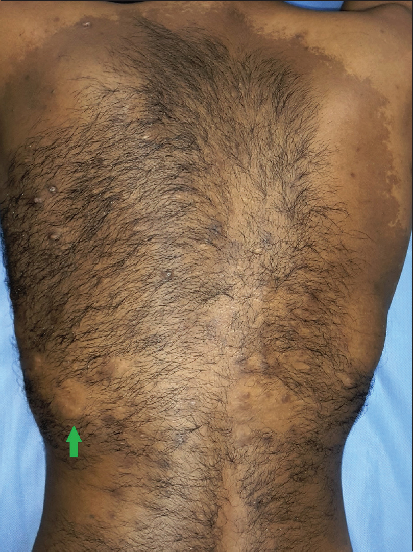 |
| Figure 1: Large brown macule with splashed geographic margin, involving almost the entire back, with increased coarse terminal hair and multiple acneiform papules. Mid-back showing an ill-defined elevated soft plaque (green arrow) |
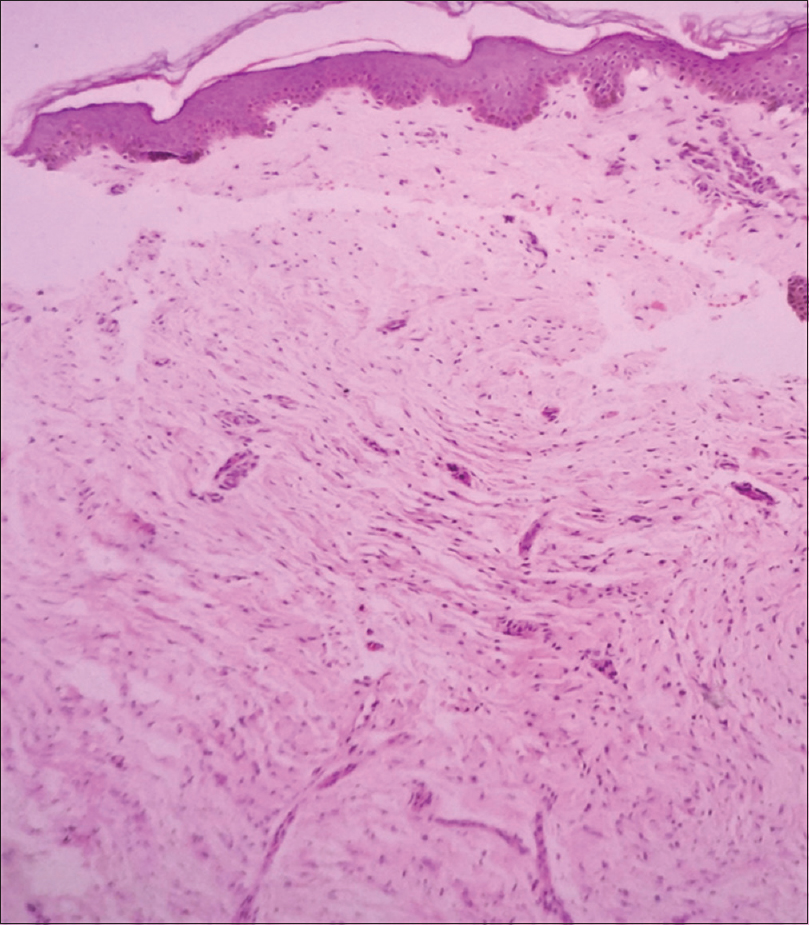 |
| Figure 2a: Biopsy from mid-back plaque showing involvement of upper, mid and deep dermis in form of spindle cell proliferation (H and E, ×100) |
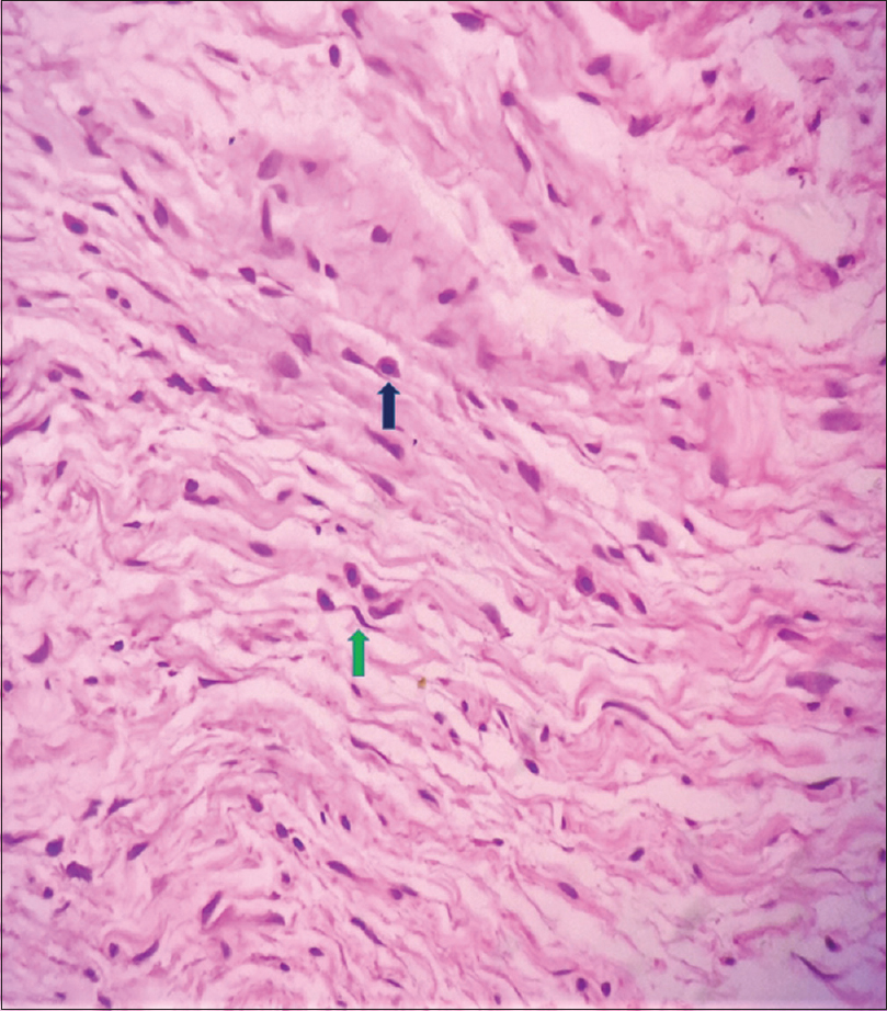 |
| Figure 2b: Biopsy from mid-back plaque showing involvement of upper, mid and deep dermis in form of spindle cell proliferation with slender, thin, wavy, tapering nuclei (green arrow) and multiple mast cells interspersed between spindle cells (blue arrow) (H and E, ×400) |
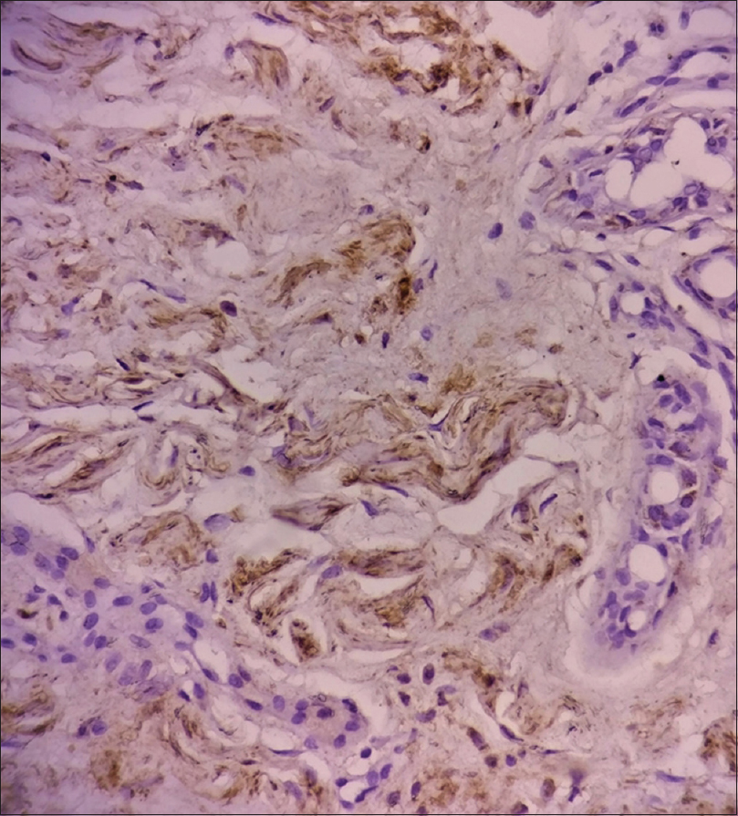 |
| Figure 3: Biopsy from mid-back plaque showing spindle cells with strong S100 staining in the mid and deep dermis (S100, ×400) |
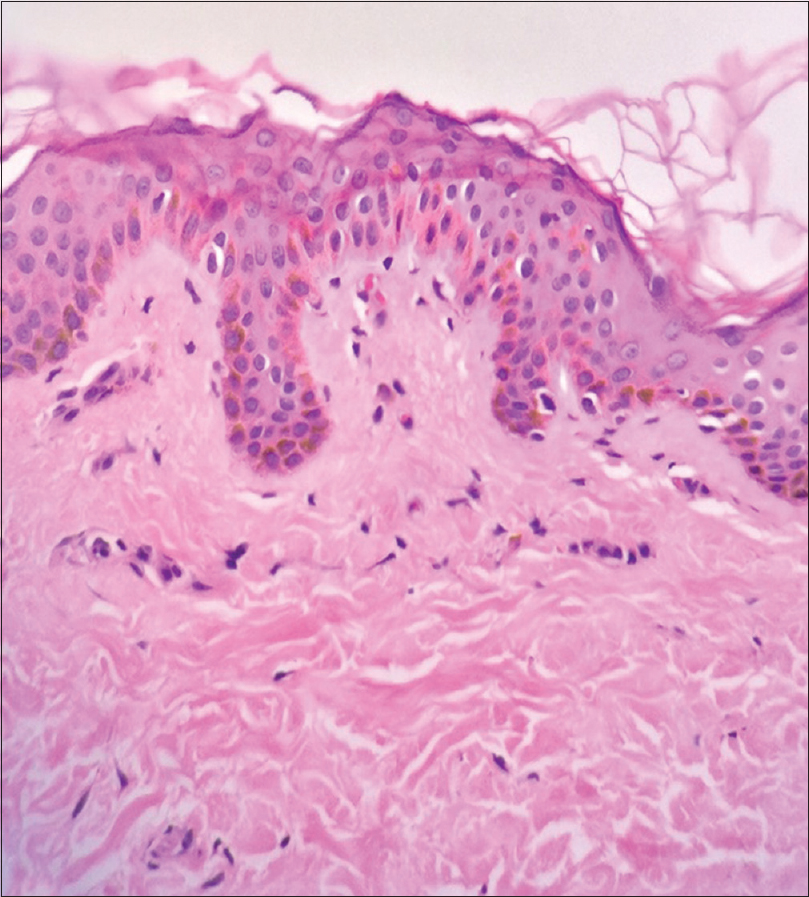 |
| Figure 4: Biopsy from the hyperpigmented macule showing elongated and squared rete ridges with basal layer hyperpigmentation (H and E, ×400) |
Becker's nevus also known as Becker's melanosis, Becker's pigmentary hamartoma, or pigmented hairy epidermal nevus is a cutaneous hamartomatous disorder characterized by a hyperpigmented macule with irregular “splashed” margins and overlying hypertrichosis. It is commonly present on the upper trunk with a prevalence of 0.5% in the general population as observed in a large French study.[1] The role of androgens have been elucidated in the pathogenesis of Becker's nevus, as evidenced by its male preponderance, peripubertal development, overlying hypertrichosis, and occasional acneiform lesions, and rare association with accessory scrotum and breast hypoplasia.[1],[2] Only few cases of congenital Becker's nevus have been described in the literature, as was seen in our case, although hypertrichosis developed during puberty.[3]
Several dermatoses occur within Becker's nevus that includes smooth muscle hamartoma, malignant melanoma, melanocytic and connective tissue nevi, perforating folliculitis, leiomyoma, lymphangioma, and very rarely, neurofibromas.[4],[5],[6] As far as ascertained, only 6 cases of Becker's nevus overlying a neurofibroma have been described in the literature.[5],[7],[8],[9],[10] Most of these were cases of neurofibromatosis type I.
Neurofibromatosis is associated with various pigmentary disorders including café au lait macules, axillary freckles, nevus of Ota, nevus spilus, congenital melanocytic nevus, and segmental unilateral melanosis.[5] Histopathology of café-au-lait macule associated with neurofibroma show increased number of functionally active melanocytes with giant melanosomes. Pigmented plexiform neurofibroma is a well-known entity in which hyperpigmentation and hypertrichosis overlying neurofibroma is seen. However, histopathology of such lesion is distinctive as it shows melanin-laden cells with spindled, dendritic, tadpole-shaped, or epithelioid morphology distributed in dermis and subcutis probably of Schwann cell origin.[11],[12] In fact, a hyperpigmented hypertrichotic flat macule is a clue to the presence of an underlying neurofibroma that may be only detected on a skin biopsy. Giant congenital melanocytic nevi could be another clinical possibility but its histopathology is distinctive and classically shows the presence of nevus cells extending into deeper dermis and subcutaneous tissue in perivascular and periadnexal location. Nevus cells can be seen in between the collagen bundles in Indian file appearance.[13] Ours was a case of neurofibroma with overlying Becker's nevus and not a pigmented plexiform neurofibroma or a giant congenital melanocytic nevi in view of the characteristic clinical finding of splashed or a geographic margin and histology showing elongated and squared hyperpigmented rete ridges suggesting Becker's nevus. Several hypothesis have been proposed to explain the association of Becker's nevus with neurofibromatosis. Fibroblasts of neurofibroma secrete several melanogenic cytokines, i.e. nerve growth factor, hepatocyte growth factor, and stem cell factor that might lead to epidermal pigmentation.[5] Moreover, mast cells secrete proopiomelanocortin and α-melanocyte stimulating hormone (α-MSH), which might also contribute to pigmentation overlying neurofibroma. Association of these two disorders existing together appears more than merely a co-incidence as both are hamartomatous proliferation disorders with increased expression of steroid hormone receptors. Molecular studies with large sample size may unfold this mystery in future.
In literature we were unable to find any previous reports of giant congenital Becker's nevus with marked hypertrichosis and multiple acneiform papules overlying a plexiform neurofibroma without any other features suggestive of neurofibromatosis type I.
Financial support and sponsorship
Nil.
Conflicts of interest
There are no conflicts of interest.
| 1. |
Grande Sarpa H, Harris R, Hansen CD, Callis Duffin KP, Florell SR, Hadley ML, et al. Androgen receptor expression patterns in Becker's nevi: An immunohistochemical study. J Am Acad Dermatol 2008;59:834-8.
[Google Scholar]
|
| 2. |
Kim YJ, Han JH, Kang HY, Lee ES, Kim YC. Androgen receptor overexpression in Becker nevus: Histopathologic and immunohistochemical analysis. J Cutan Pathol 2008;35:1121-6.
[Google Scholar]
|
| 3. |
Rao AG. Bilateral symmetrical congenital giant Becker's nevus: A Rare presentation. Indian J Dermatol 2015;60:522.
[Google Scholar]
|
| 4. |
Gupta S, Gupta S, Aggarwal K, Jain VK. Becker nevus with vitiligo and lichen planus: Cocktail of dermatoses. N Am J Med Sci 2010;2:333-5.
[Google Scholar]
|
| 5. |
Kim SY, Kim MY, Kang H, Kim HO, Park YM. Becker's naevus in a patient with neurofibromatosis. J Eur Acad Dermatol Venereol 2008;22:394-5.
[Google Scholar]
|
| 6. |
Dasegowda SB, Basavaraj G, Nischal K, Swaroop M, Umashankar N, Swamy SS, et al. Becker's nevus syndrome. Indian J Dermatol 2014;59:421.
[Google Scholar]
|
| 7. |
Kar S, Preetha K, Yadav N, Madke B, Gangane N. Becker's nevus with neurofibromatosis type 1. Ann Indian Acad Neurol 2015;18:90-2.
[Google Scholar]
|
| 8. |
Akyol M, Ozçelik S, Marufihah M, Elagöz S. Elephantiasis neuromatosa and Becker's melanosis. J Dermatol 1999;26:396-8.
[Google Scholar]
|
| 9. |
Chapel TA, Tavafoghi V, Mehregan AH, Gagliardi C. Becker's melanosis: An organoid hamartoma. Cutis 1981;27:405-6, 410, 415.
[Google Scholar]
|
| 10. |
Kim MS, Jung JY, Cho EB, Park EJ, Kim KH, Kim KJ, et al. Neurofibromas arising from Becker naevus. Clin Exp Dermatol 2015;40:814-5.
[Google Scholar]
|
| 11. |
Inaba M, Yamamoto T, Minami R, Ohbayashi C, Hanioka K. Pigmented neurofibroma: Report of two cases and literature review. Pathol Int 2001;51:565-9.
[Google Scholar]
|
| 12. |
Fetsch JF, Michal M, Miettinen M. Pigmented (melanotic) neurofibroma: A clinicopathologic and immunohistochemical analysis of 19 lesions from 17 patients. Am J Surg Pathol 2000;24:331-43.
[Google Scholar]
|
| 13. |
Viana AC, Gontijo B, Bittencourt FV. Giant congenital melanocytic nevus. An Bras Dermatol 2013;88:863-78.
[Google Scholar]
|
Fulltext Views
5,024
PDF downloads
3,111


