Translate this page into:
Giant facial cutaneous tuberculosis
2 Jinzhong City Hospital, Jinzhong City, Shanxi, China
Correspondence Address:
Jing Jun Zhao
Department of Dermatology, Tongji Hospital of Tongji University, 389, Xincun Rd, Putuo District, 200065, Shanghai
China
| How to cite this article: Ng How Tseung KS, Zhao JJ, Jia YR, Keyal U, Bhatta AK. Giant facial cutaneous tuberculosis. Indian J Dermatol Venereol Leprol 2015;81:95 |
Sir,
We report an elderly woman with facial cutaneous tuberculosis of about 30 years duration.
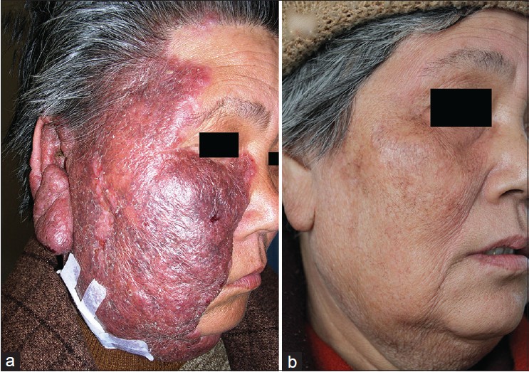
A 62-year-old woman, formerly working as a farmer in Shanxi province in China, complained of a 30-year history of a non-healing large plaque on her right face. The patient recalled that her right face was scratched 30 years ago and self-healed; two weeks later, she noticed a red lesion on the skin over the same area. The lesion, which was accompanied by mild itching and pain, gradually increased in size and became harder, eventually involving her entire right face, She had no previous records of medical examination or treatment. There was no known history of active tuberculosis in the past or of any contact with tuberculosis. She had no other illnesses. Dermatological examination revealed a well-marginated, edematous, reddish brown plaque of 28 × 18 cm in size, with a rough surface, irregular edges and mild scaling [[Figure - 1]a]. Laboratory results showed no abnormalities in blood count, liver and kidney function, blood glucose, erythrocyte sedimentation rate and urine test. Chest X-ray was normal. Tuberculin test was positive with an induration of more than 10 mm. Skin biopsy revealed a granulomatous infiltration in the dermis characterized by a central aggregation of epithelioid cells ringed by a dense collar of fibroblasts and lymphocytes; Langerhans giant cells were scattered throughout the granuloma. There were foci of caseation necrosis within the granuloma. In view of the morphology of the lesion and biopsy findings, a diagnosis of lupus vulgaris was made. The patient received antituberculous therapy having a combination of isoniazid 300 mg, rifampicin 450 mg and ethambutol 750 mg, every morning for a period of 8 weeks followed by 4 months of rifampicin and isoniazid. She was also given a topical 5% rifampicin cream to apply locally. Substantial improvement was observed after 1 month of therapy and there was complete healing after 1 year with mild residual pigmentation [[Figure - 1]b].
 |
| Figure 1: (a) Large plaque of lupus vulgaris of 30 years of duration involving the right face, ear and jaw. (b) The plaque healed with post-treatment hyperpigmentation. |
Cutaneous tuberculosis is an uncommon type of extrapulmonary tuberculosis, with a prevalence of 1.5% of all cases of tuberculosis. [1] Lupus vulgaris constitutes 74% of all cutaneous tuberculosis cases. [2] Some authors believe that lupus vulgaris is acquired mainly through endogenous inoculation, however, in the current case, we hypothesized that the exogenous scratch on the face was the cause of primary inoculation. Diagnosis of cutaneous tuberculosis relies on a combination of clinical findings, tissue culture and skin biopsy. Detection of acid-fast bacilli on culture may not always yield a positive result because lupus vulgaris is paucibacillary. Histopathological examination and response to anti-tuberculosis treatment are essential for a proper diagnosis. [3]
Categorized by clinical expression, lupus vulgaris has five forms: plaque or plane form, ulcerative or mutilating form, vegetating form, hypertrophic tumor-like form and papulo-nodular form. The plaque form is the most common type and manifests as a flat plaque with irregular edges and the surface may be smooth with psoriasiform scale. Large plaques, however, tend to show thickened and hyperkeratotic edges with irregular areas of scarring. [4] In our patient, the reddish brown plaque involved the entire right face, affecting the ipsilateral ear and part of left jaw as well while in other reports, lupus vulgaris is localized with a smaller diameter. [5] Histopathological examination showed a granulomatous inflammatory response characterized by a central aggregation of epithelioid cells and lymphocytes with occasional Langerhans giant cells. Fibrosis in the dermis indicated the chronic course of the disease. The directly observed treatment, short course (DOTS) regimen recommended for the treatment of tuberculosis consists of isoniazid 5 mg/kg/day (maximum 300 mg), rifampicin 10 mg/kg (maximum 600 mg), pyrazinamide 15 to 30 mg/kg/day (maximum 2g) and ethambutol 15 to 20 mg/kg/day (maximum 1g) for 8 weeks, and isoniazid and rifampicin 3 times weekly or twice weekly for another 4 months. [6] We treated our patient with a 8-week intensive course of rifampicin 450 mg, isoniazid 300 mg and ethambutol 750 mg administered daily, followed by 4 months of rifampicin and isoniazid in the same doses. In addition, we used a topical 5% rifampicin cream, as is the practice in China. The lesion improved within 1 month and healed completely after 1 year leaving a slight pigmentation. A 3-year post-treatment follow-up showed no recurrences.
| 1. |
Pandhi D, Reddy BS, Chowdhary S, Khurana N. Cutaneous tuberculosis in Indian children: The importance of screening for involvement of internal organs. J Eur Acad Dermatol Venereol 2004;18:546-51.
[Google Scholar]
|
| 2. |
Bhandare CA, Barad PS. Lupus vulgaris with endophthalmitis-A rare manifestation of extrapulmonary in India. Indian J Tuberc 2010;57:98-101.
[Google Scholar]
|
| 3. |
Ho CK, Ho MH, Chong LY. Cutaneous tuberculosis in Hongkong: An update. Hong Kong Med J 2006;12:272-7.
[Google Scholar]
|
| 4. |
Yates VM. Mycobacterial infections. In: Burns T, Breathnach S, Cox N, Griffiths C, editors. Rook's Textbook of Dermatology. 8 th ed. West Sussex: Wiley-Blackwell Publications; 2010. p. 31.16-9.
th ed. West Sussex: Wiley-Blackwell Publications; 2010. p. 31.16-9.'>[Google Scholar]
|
| 5. |
Sacchidanand S, Sharavana S, Mallikarjun M, Nataraja HV. Giant lupus vulgaris: A rare presentation. Indian Dermatol Online J 2012;3:34-6.
[Google Scholar]
|
| 6. |
Handog EB, Gabriel TG, Pineda RT. Management of cutaneous tuberculosis. Dermatol Ther 2008;21:154-61.
[Google Scholar]
|
Fulltext Views
4,030
PDF downloads
3,048


