Translate this page into:
Giant proliferating trichilemmal tumour of the scalp
Corresponding author: Dr. Sang-Yeul Lee, Department of Plastic and Reconstructive Surgery, Kangwon National University Hospital, Baekryeong-ro 156, Chuncheon, Kangwon-do, Republic of Korea. serafin5@unitel.co.kr
-
Received: ,
Accepted: ,
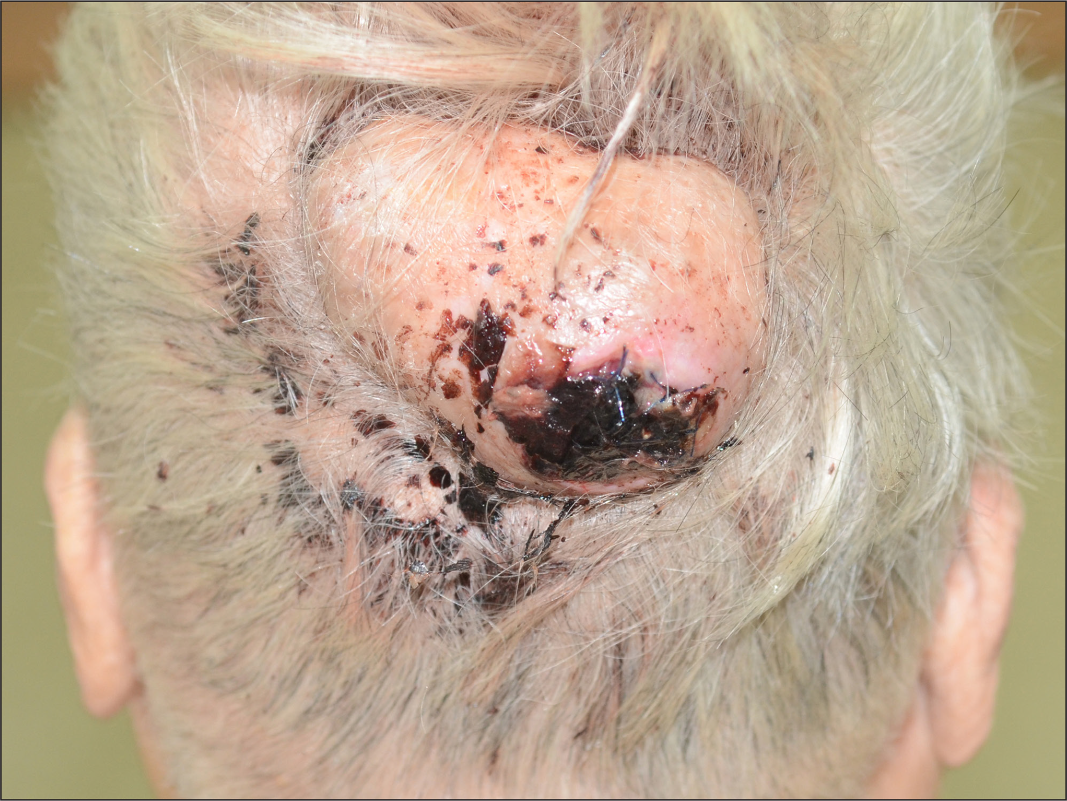
An 88-year-old woman presented with a large, asymptomatic, slow-growing scalp mass that had been persisting for over 40 years. Physical examination revealed a flesh-coloured, dome-shaped mass with ulceration and haemorrhagic crusting in the occipital region, measuring 8 × 5 × 3.5 cm [Figure 1]. The tumour was resected with a 10 mm margin of normal scalp tissue over the pericranium. Histopathologically, the lesion showed squamous epithelial proliferation and trichilemmal keratinization, with no evidence of malignancy or local invasion. Proliferating trichilemmal tumours are uncommon adnexal neoplasms frequently occurring on the scalp, usually benign but with locally invasive and even metastatic potential. Abnormally large proliferating trichilemmal tumours, described as giant proliferating trichilemmal tumours, commonly have long clinical histories and develop skin changes such as ulceration and necrosis. The clinical features that predict a risky prognosis include a rapid increase in size, tumour size greater than 5 cm, non-scalp location and foci of necrosis or ulceration. Complete surgical excision of the tumour with a 1 cm margin of normal tissue is the proper therapy to prevent local recurrence.

A flesh-coloured, dome-shaped mass (8 × 5 × 3.5 cm) with ulceration and haemorrhagic crusting in the occipital region
Declaration of patient consent
The authors certify that they have obtained all appropriate patient consent.
Financial support and sponsorship
Nil.
Conflicts of interest
There are no conflicts of interest.


