Generic selectors
Exact matches only
Search in title
Search in content
Post Type Selectors
Search in posts
Search in pages
Filter by Categories
15th National Conference of the IAOMFP, Chennai, 2006
Abstract
Abstracts from current literature
Acne in India: Guidelines for management - IAA Consensus Document
Addendum
Announcement
Art & Psychiatry
Article
Articles
Association Activities
Association Notes
Award Article
Book Review
Brief Report
Case Analysis
Case Letter
Case Letters
Case Notes
Case Report
Case Reports
Clinical and Laboratory Investigations
Clinical Article
Clinical Studies
Clinical Study
Commentary
Conference Oration
Conference Summary
Continuing Medical Education
Correspondence
Corrigendum
Cosmetic Dermatology
Cosmetology
Current Best Evidence
Current Issue
Current View
Derma Quest
Dermato Surgery
Dermatopathology
Dermatosurgery Specials
Dispensing Pearl
Do you know?
Drug Dialogues
e-IJDVL
Editor Speaks
Editorial
Editorial Remarks
Editorial Report
Editorial Report - 2007
Editorial report for 2004-2005
Errata
Erratum
Focus
Fourth All India Conference Programme
From Our Book Shelf
From the Desk of Chief Editor
General
Get Set for Net
Get set for the net
Guest Article
Guest Editorial
History
How I Manage?
IADVL Announcement
IADVL Announcements
IJDVL Awards
IJDVL AWARDS 2015
IJDVL Awards 2018
IJDVL Awards 2019
IJDVL Awards 2020
IJDVL International Awards 2018
Images in Clinical Practice
Images in Dermatology
In Memorium
Inaugural Address
Index
Knowledge From World Contemporaries
Leprosy Section
Letter in Response to Previous Publication
Letter to Editor
Letter to the Editor
Letter to the Editor - Case Letter
Letter to the Editor - Letter in Response to Published Article
LETTER TO THE EDITOR - LETTERS IN RESPONSE TO PUBLISHED ARTICLES
Letter to the Editor - Observation Letter
Letter to the Editor - Study Letter
Letter to the Editor - Therapy Letter
Letter to the Editor: Articles in Response to Previously Published Articles
Letters in Response to Previous Publication
Letters to the Editor
Letters to the Editor - Letter in Response to Previously Published Articles
Letters to the Editor: Case Letters
Letters to the Editor: Letters in Response to Previously Published Articles
Media and news
Medicolegal Window
Messages
Miscellaneous Letter
Musings
Net Case
Net case report
Net Image
Net Images
Net Letter
Net Quiz
Net Study
New Preparations
News
News & Views
Obituary
Observation Letter
Observation Letters
Oration
Original Article
ORIGINAL CONTRIBUTION
Original Contributions
Pattern of Skin Diseases
Pearls
Pediatric Dermatology
Pediatric Rounds
Perspective
Presedential Address
Presidential Address
Presidents Remarks
Quiz
Recommendations
Regret
Report
Report of chief editor
Report of Hon : Treasurer IADVL
Report of Hon. General Secretary IADVL
Research Methdology
Research Methodology
Resident page
Resident's Page
Resident’s Page
Residents' Corner
Residents' Corner
Residents' Page
Retraction
Review
Review Article
Review Articles
Reviewers 2022
Revision Corner
Self Assessment Programme
SEMINAR
Seminar: Chronic Arsenicosis in India
Seminar: HIV Infection
Short Communication
Short Communications
Short Report
Snippets
Special Article
Specialty Interface
Studies
Study Letter
Study Letters
Supplement-Photoprotection
Supplement-Psoriasis
Symposium - Contact Dermatitis
Symposium - Lasers
Symposium - Pediatric Dermatoses
Symposium - Psoriasis
Symposium - Vesicobullous Disorders
SYMPOSIUM - VITILIGO
Symposium Aesthetic Surgery
Symposium Dermatopathology
Symposium-Hair Disorders
Symposium-Nails Part I
Symposium-Nails-Part II
Systematic Review and Meta-Analysis
Systematic Reviews and Meta-analyses
Systematic Reviews and Meta-analysis
Tables
Technology
Therapeutic Guideline-IADVL
Therapeutic Guidelines
Therapeutic Guidelines - IADVL
Therapeutics
Therapy
Therapy Letter
Therapy Letters
View Point
Viewpoint
What’s new in Dermatology
View/Download PDF

Translate this page into:

Letter to the Editor
2011:77:4;517-519
doi: 10.4103/0378-6323.82391
PMID: 21727705

Gigantic cutaneous rhinosporidiosis with giant cells bloated with sporangia

Ashok K Ghorpade
 Department of Dermatology and Venereology, J. L. N. Hospital and Research Centre, Bhilai Steel Plant, Bhilai, Chhattisgarh, India

Correspondence Address:
Ashok K Ghorpade
BK D-18, Sector 9, Bhilai, Chhattisgarh - 490 006
India
How to cite this article:
Ghorpade AK. Gigantic cutaneous rhinosporidiosis with giant cells bloated with sporangia. Indian J Dermatol Venereol Leprol 2011;77:517-519
Copyright: (C)2011 Indian Journal of Dermatology, Venereology, and Leprology

Sir,

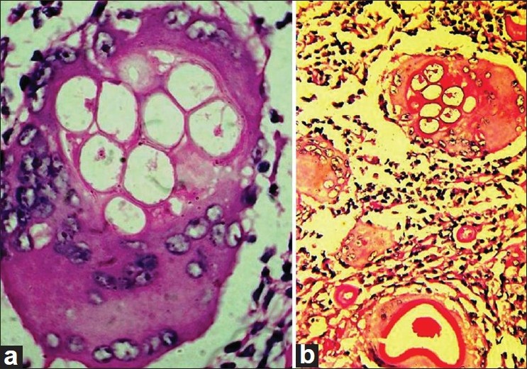
A 21-year-old Indian male presented with multiple, asymptomatic skin lesions on his left leg and foot of 18 months duration. He was operated upon for a nasal growth causing bleeding and some globoid skin lesions on his left calf by a private doctor about 3 months earlier without histopathological examination. There was history of bathing in the common village pond along with buffaloes, since early childhood. There was no family history of skin or nasal lesions. Cutaneous examination showed skin colored, firm globoid skin lesion about 5 × 5 × 5 cm 3 in size on the dorsal aspect of the left foot and left leg, the former with a papular lesion about 3 mm in diameter near its base [Figure - 1]a and similar smaller ones on posterior aspect of the left leg with scars of excised lesions and varicose veins [Figure - 1]b. There were no nasal lesions, organomegaly, or lymphadenopathy at the time of examination. Systemic examination was normal. Routine hematology, blood glucose, liver and kidney function tests, abdominal ultrasound, and X-ray of the limbs were normal and ELISA for HIV 1 and 2 was negative. Histopathology from the globoid skin lesion showed foreign body giant cells bloated with engulfed sporangia and multiple sporangia with endospores in the dermis with a chronic inflammatory infiltrate [Figure - 2]a and b. Crushed tissue smear of the papular lesion also revealed similar sporangia with endospores. The smaller skin lesions were excised followed by electrocoagulation of the base, and he was given tablet dapsone 100 mg once daily without any recurrence during the follow-up of 4 months. He was advised excision for the bigger lesions.
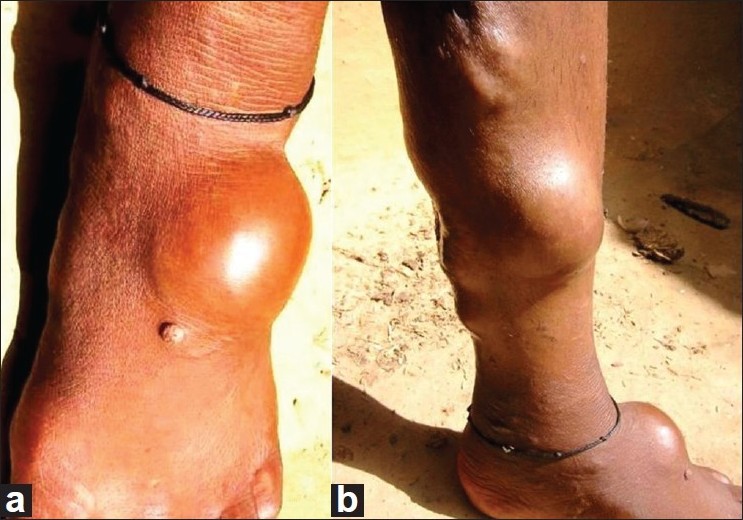
Figure 1: (a) Co-existing big globoid lesion and a papule on the left foot. (b) Multiple, huge, and small globoid skin lesions on the left leg with scars of excised lesions and varicose veins
Figure 2: (a) Close-up of a foreign body giant cell seen bloated with sporocysts in the upper half, and the nuclei pushed to edges (hematoxylin and eosin; original magnification, ×1000). (b) Same foreign body giant cell (as in (a)) in the upper part, with another one on the lower end. Thick-walled sporocysts with pinkish nutritional material in the lower and upper edges, two foreign body giant cells in the middle and a dense chronic inflammatory infiltrate (Periodic acid Schiff stain; original magnification, ×400)

Rhinosporidiosis is endemic in Sri Lanka and parts of India but has been reported from other parts namely Africa, South America, and the United States. In India, Chhattisgarh, Tamil Nadu, Kerala, Karnataka, Pondicherry, and Orissa are endemic for the disease. Several cases of nasal and cutaneous rhinosporidiosis have been described from the Chhattisgarh state. [1],[2],[3],[4] In addition, sporadic cases have been described among migrant population, from other parts as well. Most of the patients are young/middle-aged males, and sharing pond bathing with cattle and repeated trauma seem to be the predisposing factors here. [1],[2],[3],[4] Rhinosporidiosis is caused by Rhinosporidium seeberi, an aquatic protistan parasite of the Mesomycetozoea class along with organisms causing similar infections in amphibians and fish. It is the first known human pathogen from the DRIPs clade. [5] There are no reports of successful in vitro culture and propagation in subculture with various types of culture media. The disease may also affect farm, domestic, and wild animals. It is commoner in males between the second to fourth decades probably due to the increased chances of exposure. The exact mode of transmission is unclear, but it might be acquired through contaminated dust, water, infected clothing, or fingers. There is mucosal involvement of nose, nasopharynx, and soft palate in the form of sessile or pedunculated vascular polyps, studded with minute whitish dots (sporangia containing the spores). Nasal obstruction and bleeding are common. Occasionally, it involves the lips, palate, uvula, maxillary antrum, epiglottis, larynx, trachea, bronchus, ear, scalp, vulva, vagina, penis, rectum, and the skin. Cutaneous rhinosporidiosis may occur from the nasal growths as satellite lesions, or as disseminated lesions with or without nasal involvement, or as primary skin lesions. They begin as papules or nodules and may spread by scratching, inoculating the organism with contaminated fingers from nasal picking, through blood or the lymphatics. [3],[4] They may resemble verruca vulgaris, tuberculosis verrucosa cutis, granuloma pyogenicum, venereal warts, or donovanosis. Cutaneous lesions at sites distant from the nasal mucosa are uncommon.

A clinical diagnosis of gigantic cutaneous rhinosporidiosis with co-existing globoid and papular skin lesions was made. Lipomas, liposarcomas, and subcutaneous aspergillosis were considered in the differential diagnosis, but the history of a nasal growth and bleeding, shared pond bathing with buffaloes, the firm globoid lesions associated with the papular skin lesions, the earlier experience of dealing with such cases suggested and histopathology confirmed the diagnosis. Coccidio idomycosis has a different clinical presentation and a smaller sporangia size. Disseminated polymorphic cutaneous rhinosporidiosis, with verrucoid, furunculoid, and a pseudohorn-like skin lesion was reported by Kumari et al. [6] Nayak et al. [7] described similar but disseminated subcutaneous globoid lesions along with sessile and pedunculated moist, shiny grouped and discrete, highly vascular swellings all over the body and scalp. Although such giant cells bloated with sporangia have been reported once in a patient having a single subcutaneous lesion and nasal rhinosporidiosis, [3] they have not been observed in a patient with polymorphic cutaneous lesions.

Treatment is by excision with electrocoagulation, but recurrences may occur. Oral dapsone might arrest the maturation of the sporangia and cause stromal fibrosis. [8]

References
1.
Sudarshan V, Goel NK, Gahine R, Krishnani C. Rhinosporidiosis in Raipur, Chhattisgarh: A report of 462 cases. Indian J Pathol Microbiol 2007;50:718-21.
[Google Scholar]
2.
Ramanan C, Ghorpade A. Giant cutaneous rhinosporidiosis. Int J Dermatol 1996;35:441-2.
[Google Scholar]
3.
Ghorpade A. Rhinosporidiosis: Gigantic cells with engulfed sporangia of Rhinospoirdia seeberi in a case of dermosporidiosis. Int J Dermatol 2008;47:694-5.
[Google Scholar]
4.
Acharya PV, Gupta RL, Darbari BS. Cutaneous rhinosporidiosis. Indian J Dermatol Venereol Leprol 1973;39:22-5.
[Google Scholar]
5.
Fredricks DN, Jolley JA, Lepp PW, Kosek JC, Relman DA. Rhinosporidium seeberi: A human pathogen from a novel group of aquatic protistan parasites. Emerg Infect Dis 2000;6:273-82.
[Google Scholar]
6.
Kumari R, Nath AK, Rajalakshmi R, Adityan B, Thappa DM. Disseminated cutaneous rhinosporidiosis: Varied morphological appearances on the skin. Indian J Dermatol Venereol Leprol 2009;75:68-71.
[Google Scholar]
7.
Nayak S, Acharjya B, Devi B, Sahoo A, Singh N. Disseminated cutaneous rhinosporidiosis. Indian J Dermatol Venereol Leprol 2007;73:185-7.
[Google Scholar]
8.
Thappa DM, Venkatesan S, Sirka CS, Jaisankar TJ, Gopalkrishnan, Ratnakar C. Disseminated cutaneous rhinosporidiosis. J Dermatol 1998;25:527-32.
[Google Scholar]

Fulltext Views
1,353

PDF downloads
1,369
Show Sections