Generic selectors
Exact matches only
Search in title
Search in content
Post Type Selectors
Search in posts
Search in pages
Filter by Categories
15th National Conference of the IAOMFP, Chennai, 2006
Abstract
Abstracts from current literature
Acne in India: Guidelines for management - IAA Consensus Document
Addendum
Announcement
Art & Psychiatry
Article
Articles
Association Activities
Association Notes
Award Article
Book Review
Brief Report
Case Analysis
Case Letter
Case Letters
Case Notes
Case Report
Case Reports
Clinical and Laboratory Investigations
Clinical Article
Clinical Studies
Clinical Study
Commentary
Conference Oration
Conference Summary
Continuing Medical Education
Correspondence
Corrigendum
Cosmetic Dermatology
Cosmetology
Current Best Evidence
Current Issue
Current View
Derma Quest
Dermato Surgery
Dermatopathology
Dermatosurgery Specials
Dispensing Pearl
Do you know?
Drug Dialogues
e-IJDVL
Editor Speaks
Editorial
Editorial Remarks
Editorial Report
Editorial Report - 2007
Editorial report for 2004-2005
Errata
Erratum
Focus
Fourth All India Conference Programme
From Our Book Shelf
From the Desk of Chief Editor
General
Get Set for Net
Get set for the net
Guest Article
Guest Editorial
History
How I Manage?
IADVL Announcement
IADVL Announcements
IJDVL Awards
IJDVL AWARDS 2015
IJDVL Awards 2018
IJDVL Awards 2019
IJDVL Awards 2020
IJDVL International Awards 2018
Images in Clinical Practice
In Memorium
Inaugural Address
Index
Knowledge From World Contemporaries
Leprosy Section
Letter in Response to Previous Publication
Letter to Editor
Letter to the Editor
Letter to the Editor - Case Letter
Letter to the Editor - Letter in Response to Published Article
LETTER TO THE EDITOR - LETTERS IN RESPONSE TO PUBLISHED ARTICLES
Letter to the Editor - Observation Letter
Letter to the Editor - Study Letter
Letter to the Editor - Therapy Letter
Letter to the Editor: Articles in Response to Previously Published Articles
Letters in Response to Previous Publication
Letters to the Editor
Letters to the Editor - Letter in Response to Previously Published Articles
Letters to the Editor: Case Letters
Letters to the Editor: Letters in Response to Previously Published Articles
Media and news
Medicolegal Window
Messages
Miscellaneous Letter
Musings
Net Case
Net case report
Net Image
Net Images
Net Letter
Net Quiz
Net Study
New Preparations
News
News & Views
Obituary
Observation Letter
Observation Letters
Oration
Original Article
ORIGINAL CONTRIBUTION
Original Contributions
Pattern of Skin Diseases
Pearls
Pediatric Dermatology
Pediatric Rounds
Perspective
Presedential Address
Presidential Address
Presidents Remarks
Quiz
Recommendations
Regret
Report
Report of chief editor
Report of Hon : Treasurer IADVL
Report of Hon. General Secretary IADVL
Research Methdology
Research Methodology
Resident page
Resident's Page
Resident’s Page
Residents' Corner
Residents' Corner
Residents' Page
Retraction
Review
Review Article
Review Articles
Reviewers 2022
Revision Corner
Self Assessment Programme
SEMINAR
Seminar: Chronic Arsenicosis in India
Seminar: HIV Infection
Short Communication
Short Communications
Short Report
Snippets
Special Article
Specialty Interface
Studies
Study Letter
Study Letters
Supplement-Photoprotection
Supplement-Psoriasis
Symposium - Contact Dermatitis
Symposium - Lasers
Symposium - Pediatric Dermatoses
Symposium - Psoriasis
Symposium - Vesicobullous Disorders
SYMPOSIUM - VITILIGO
Symposium Aesthetic Surgery
Symposium Dermatopathology
Symposium-Hair Disorders
Symposium-Nails Part I
Symposium-Nails-Part II
Systematic Review and Meta-Analysis
Systematic Reviews and Meta-analyses
Systematic Reviews and Meta-analysis
Tables
Technology
Therapeutic Guideline-IADVL
Therapeutic Guidelines
Therapeutic Guidelines - IADVL
Therapeutics
Therapy
Therapy Letter
Therapy Letters
View Point
Viewpoint
What’s new in Dermatology
View/Download PDF

Translate this page into:

Net letter
2012:78:3;409-409
doi: 10.4103/0378-6323.95495

Keloidal atypical fibroxanthoma on the back of an 80-year-old man, pitfalls in the clinical and histological diagnosis

Hsiao Yi-Hsin, Chiu Cheng-Sheng
 Department of Dermatology, Chang Gung Memorial Hospital, Taipei; Chang Gung University College of Medicine, Taoyuan, Taiwan

Correspondence Address:
Chiu Cheng-Sheng
Department of Dermatology, Chang Gung Memorial Hospital, 199, Tun Hwa North Road, Taipei 105
Taiwan
How to cite this article:
Yi-Hsin H, Cheng-Sheng C. Keloidal atypical fibroxanthoma on the back of an 80-year-old man, pitfalls in the clinical and histological diagnosis. Indian J Dermatol Venereol Leprol 2012;78:409
Copyright: (C)2012 Indian Journal of Dermatology, Venereology, and Leprology

Sir,

Keloidal atypical fibroxanthoma (AFX) is a new and under-recognized histological variant of AFX. We, herein, report a case of a keloidal AFX with the unusual presentation of a rapid growing, ulcerative plaque on the back of an 80-year-old male.

An 80-year-old man presented with an asymptomatic, rapid growing, infiltrating erythematous plaque on his back for 3 months. He denied previous trauma in this area. There was no constitutional symptoms such as fever or weight loss. Clinical examination revealed a 7 × 5 cm, ill-defined, firm and violaceous plaque on the lower back. Overlying epidermis was erosive and crusted [Figure - 1]. Lymph node examinations revealed no neck, axillary or inguinal lymphadenopathy. Other physical, examinations showed no abnormal findings. His comorbidities included Parkinsonism, hypertension and hyperlipidemia. A squamous cell carcinoma was clinically suspected and an incisional biopsy on the plaque was performed. Histopathological findings revealed a poor circumscribed, hypercellular tumor extending deeply into the reticular dermis [Figure - 2]a. The lesion revealed diffuse infiltration of pleomorphic spindle cells around prominent keloidal broad collagen fibers. The neoplastic cells demonstrated prominent atypia with bizarre, large hyper chromatic nuclei and numerous mitotic Figures [Figure - 2]b. Some larger tumor cells contained cytoplasmic hyaline globules with hemosiderin granules [Figure - 2]c. Tentative pathological diagnosis was sarcomatoid pleomorphic tumor. Immuonohistochemical study was performed for further differential diagnosis.
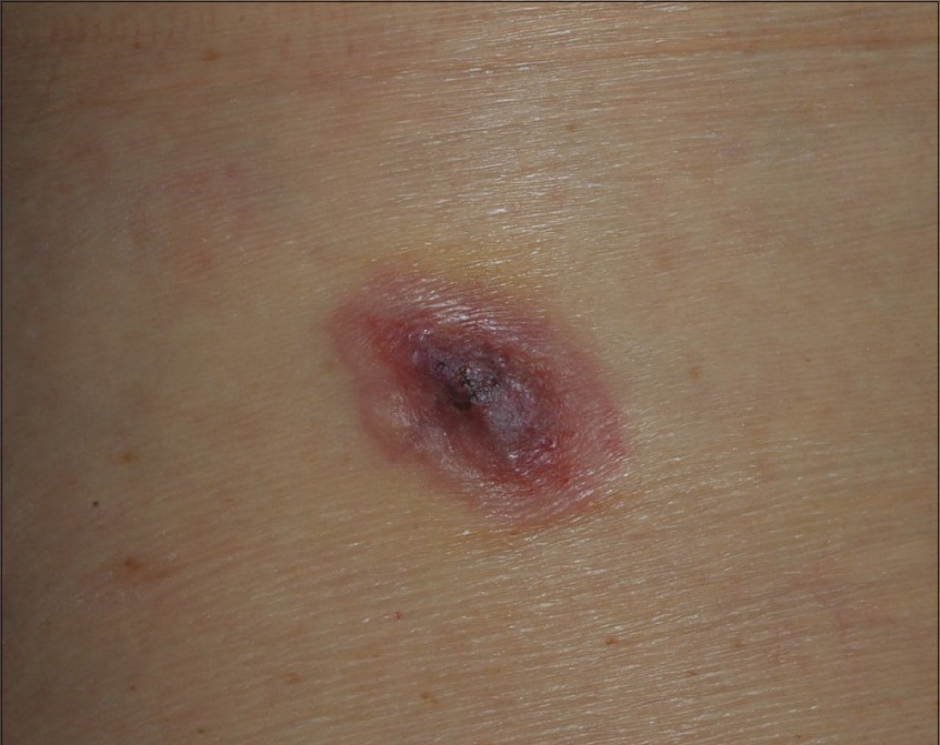
Figure 1: A 7 × 5 cm, ill-defined, erythematous to violaceous plaque on the back with surface erosion
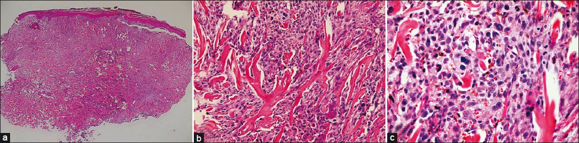
Figure 2: (a) Low power field revealed a poor circumscribed, hyper-cellular tumor extending deeply into the reticular dermis (H and E, ×40), (b) The lesion revealed diffuse infiltration of pleomorphic spindle cells around prominent keloidal broad collagen fibers. The neoplastic cells demonstrated prominent atypia with bizarre, large hyper chromatic nuclei. and numerous mitotic Figures (H and E, ×100). (c) Some larger tumor cells contained cytoplasmic hyaline globules with hemosiderin granules (H and E, ×400)
Immunohistochemical staining results showed that the tumor cells were positive for Vimentin [Figure - 3]a, CD10 [Figure - 3]b, smooth muscle actin, CD68 (KP1) and CD99. Pancytokeratin: AE1 AE3 [Figure - 3]c, S100 protein [Figure - 3]d, HMB45, CD31, factor XIIIa and desmin were negative. Diagnosis of Keloidal AFX was made based on the clinical and pathological findings. Wide excision with 1 cm surgical margin was performed. The patient has remained free of recurrence for 1-year follow-up.
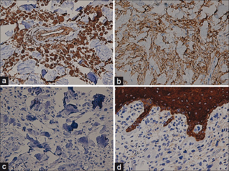
Figure 3: (a and b) Immunohistochemical features: Most spindle cells expressed Vimentin (a) and CD10 (b) (×400). (c and d) Negative S100 (c) and Pancytokeratin (d) immunohistochemical staining presented throughout the neoplasm (×400)

AFX, a pleomorphic fibrohistiocytic tumor, develops prominently on actinically damaged skin on the head and neck of elderly. Clinically, AFX begins as a rapid growing, solitary erythematous nodule and may be ulcerated. There are bimodal distribution of anatomical location and age of AFX. [1] Most of the patients are in the 7 th -8 th decade and present with AFX on the face and neck, indicating previous solar damage. Conversely, smaller group of patients present in their 4 th -5 th decade with AFX on the trunk and limbs without prominent actinic damage. [2] Our case is unique with the presentation of a keloidal AFX on the back, an uncharacteristic location for his demographic group, which raises the clinical diagnostic difficulties.

Histologically, AFX was characterized by sarcomatous appearance of mixed spindle and bizarre pleomorphic cells with evident mitoses and xanthomatous histiiocytes. [3] Histological variants of AFX included tumors with pigment, clear cell change, granular cell change, osteoclast-like giant cell and keloidal changes.

Keloidal AFX, first formally described in 2009, [3] is an unusual and under-recognized histological variant. Typical keloidal AFX is characterized by atypical pleomorphic fibrohistiocytic cells trapped within interstitial thick, glassy, eosinophilic collagen throughout the majority of the tumor. Occasionally, the keloidal collagen distribute around ring-shaped vascular structures. [4]

Clinical differential diagnosis of our case includes squamous cell carcinoma, metastatic carcinoma, melanoma or sarcoma. Histologically, pleomorphic spindle cells with keloidal collagens result in diagnostic challenges in the classical histopathological study. Further immunohistochemistry techniques are required to exclude malignant and benign neoplasm with keloidal change and dermatofibroma with monster cell. AFX typically expresses Vimentin, CD10, CD68 and CD99. Lack of S100 staining distinguishes the lesion from desmoplastic melanoma. Absence of pancytokeratin and desmin staining differentiate AFX from spindle cell squamous cell carcinoma and metastatic leiomyosarcoma, respectively. [4] Dermatofibroma with monster cell can be excluded upon negative staining of factor XIIIa.

The presence of keloidal collagen in AFX does not influence the clinical prognosis. [3] In spite of the highly atypical cytological appearance, general prognosis of AFX is good. Local recurrence is reported in 5% of the patient but metastases are exceptional. [5] Surgical excision is the main treatment strategy. In poorly circumscribed tumors with infiltrative border, excision with wider margin is suggested to avoid recurrence. Mohs surgery has been recommended by case series as a favorable method to optimize cosmetic outcome and decrease the recurrence rate. [4],[5]

With the combination of unusual clinical presentation and prominent keloidal collagen in highly anaplastic histological appearance, misdiagnosis may result in unnecessary evaluation and overextensive surgical intervention. This case highlights the integration of clinical, histological and immunohistochemical features to correctly diagnose keloidal AFX. Regardless of lesion location and patient age, keloidal AFX should always be taken into differential consideration when encountering plemorphic spindle cell tumors containing keloidal collagen.

References
1.
Fussell JN, Cooke ER, Florentino F, Sheehan DJ. Atypical fibroxanthoma with keloidal collagen. Am J Dermatopathol 2010;32:713-5.
[Google Scholar]
2.
Kim J, McNiff JM. Keloidal atypical fibroxanthoma: A case Series. J Cutan Pathol 2009;36:535-9.
[Google Scholar]
3.
Offman S, Pasternak S, Walsh N. Keloidal and other collagen patterns in atypical fibroxanthomas. Am J Dermatopathol 2010;32:326-32.
[Google Scholar]
4.
Davis JL, Randle HW, Zalla MJ, Roenigk RK, Brodland DG. A comparison of Mohs micrographic surgery and wide excision for the treatment of atypical fibroxanthoma. Dermatol Surg 1997;23:105-10.
[Google Scholar]
5.
Cooper JZ, Newman SR, Scott GA, Brown MD. Metastasizing atypical fibroxanthoma (cutaneous malignant histiocytoma): Report of five cases. Dermatol Surg 2005;31:221-5.
[Google Scholar]

Fulltext Views
921

PDF downloads
995
Show Sections