
Translate this page into:
Kissing lesions in dermatology
Corresponding author: Dr. Prakhar Srivastava, Department of Dermatology & STD, Pandit Madan Mohan Malaviya Hospital, Delhi, India. doctorprakharsjh@gmail.com
-
Received: ,
Accepted: ,
How to cite this article: Srivastava P, Pulumati A, Dash A, Khunger N. Kissing lesions in dermatology. Indian J Dermatol Venereol Leprol. 2025;91:271-4. doi: 10.25259/IJDVL_26_2024
Introduction
There are numerous differential diagnoses for “Kissing lesions” in dermatology.1 It is imperative also to differentiate kissing lesions from flexural dermatoses. Flexural dermatoses refer to skin conditions that primarily affect skin folds such as inverse psoriasis, intertrigo, atopic dermatitis, and Hailey-Hailey disease. These are usually generalised in distribution and may involve a large body surface area.2 In contrast, kissing lesions describe a specific pattern where two small, discrete, well-defined lesions/ sets of lesions occur in mirror image locations on opposing skin surfaces. Flexural dermatoses and kissing lesions are terms that are used to describe different sets of skin conditions, but these are not mutually exclusive; a skin disorder may exhibit both flexural involvement and kissing lesions, depending on its nature and presentation.
1. Congenital
The ‘divided’ or ‘kissing’ naevus was first reported by Van Michael Paul in 1908 and named by Fuchs in 1919. Fuchs described a congenital melanocytic naevus on adjacent parts of the upper and lower eyelid giving the appearance of a single lesion when the eye was closed. Kissing or divided naevus can only be seen on those parts of the body at which separation occurs during embryogenesis.3 Because this separation occurs after melanoblast migration to the skin, the resulting pigmented lesion appears to be divided in two. Some of the commonly involved sites include upper and lower eyelids separated by palpebral fissure and inner foreskin and dorsal glans penis separated by coronal sulcus.
-
A.
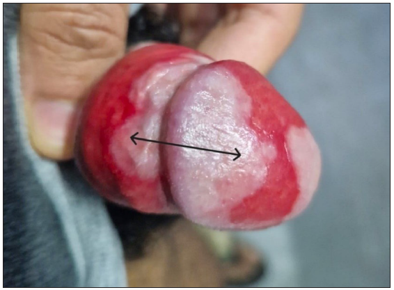
Kissing or divided congenital melanocytic naevus – In rare cases, melanocytic naevi may exhibit lesions in a mirror image configuration or kissing pattern. They have been reported to occur on eyelids, penis [Figure 1], lips, and labia majora.4–6 Kissing naevus of eyelids has also been called as ‘split ocular naevus’ or ‘panda naevus’.7
-
B.
Divided spotted grouped pigmented naevus – On upper and lower eyelids.8
-
C.
Divided naevus spilus – On upper and lower eyelids.8
-
D.
Divided epidermal naevus – On adjacent fingers.9
-
E.
Divided mastocytoma – Described as divided mast cell naevus located on adjacent fingers.10
-
F.
Kissing café au lait macules – On upper and lower eyelids.11
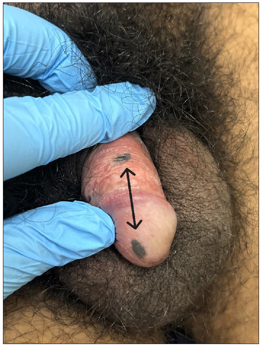
- Kissing melanocytic naevus of the penis.
2. Infections
Non-sexually transmitted
-
A.
Oral candidiasis – Median rhomboid glossitis on the tongue has been known to occur along with kissing palatal candidiasis. The kissing lesion on the palate is characterised by erythema resulting from contact with the tongue.12
-
B.
Impetigo, Intertrigo – These are usually described as flexural dermatoses which may in some situations also have kissing lesions.
Sexually transmitted infections
-
A.
-
B.
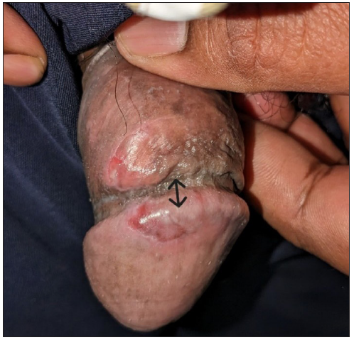
Kissing chancroid – Kissing chancroid can occur on the glans and adjacent inner foreskin [Figure 3].
-
C.
Kissing condyloma lata – Condyloma lata, a manifestation of secondary syphilis, typically present as raised gray-white lesions on mucosal areas. These lesions often have a wart-like appearance and can occur in the genital and anal regions and often demonstrate a kissing morphology.14
-
D.
Genital warts and molluscum contagiosum: These may present as kissing lesions due to pseudo-koebnerisation.
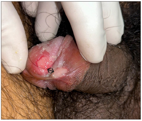
- Kissing chancre (resolving) of the penis.
- Kissing chancroid of the penis.
3. Irritant
-
A.
Paederus dermatitis (blister beetle dermatitis or dermatitis linearis) – It presents with an intensely burning linear or serpiginous rash in a kissing or mirror image pattern [Figure 4]. The kissing pattern is caused by the crushing of the beetle against two flexor surfaces, causing a release of Pederin toxin (which is found in the hemolymph of Paederus) onto the skin.15
-
B.
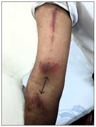
- Kissing paederus dermatitis on the right upper limb.
- Kissing Zoon’s balanitis.
4. Inflammatory and Metabolic
-
A.
Lipschütz ulcer – This is a rare and acute form of genital ulcers that primarily affects young, sexually active women. While it was initially described in the context of infectious mononucleosis, these ulcers can occur in the absence of this infection. Other factors, such as viral or bacterial infections, systemic illnesses, and hypersensitivity reactions, have been suggested as potential triggers. The condition is characterized by the sudden appearance of painful, shallow ulcers on the labia minora, vestibule, or perineum. The ulcers are typically solitary or few, with well-defined borders. They may be associated with systemic symptoms such as fever and malaise.17
-
B.
Kissing hidradenitis suppurativa – They have been described on the penile shaft and opposing scrotum.1
-
C.
Porokeratosis ptychotropica – It is a disorder of keratinization clinically characterised by a symmetrical reddish to brown-coloured hyperkeratotic, verrucous or psoriasiform plaques on the perianal and gluteal regions in a kissing-like or butterfly pattern.18
-
D.
Kissing xanthelasma – There is only one documented case of kissing xanthelasma in a five-year-old girl with unilateral kissing xanthelasma on the upper and lower left eyelids without abnormal serum lipid levels.19
5. Neoplastic
-
A.
Kissing basaloid follicular hamartoma – Basaloid follicular hamartoma is a rare benign neoplasm that typically presents as multiple small papules or nodules on the skin. They have been reported to occur in a kissing fashion over the upper and lower eyelids.20
-
B.
Kissing melanoma – This rare entity was reported on the prepuce and glans of a 30-year-old male arising in a pre-existing kissing melanocytic naevus of the penis.21
-
C.
Kissing sebaceous carcinoma – This has been reported on upper and lower eyelids. The kissing tumours might have independently developed in the adjacent eyelids, or one tumour appeared first and then invaded to the neighbouring eyelid through pagetoid spreading in a continuous way.22
Some of the well-described causes of kissing lesions have been summarised in Figure 6.
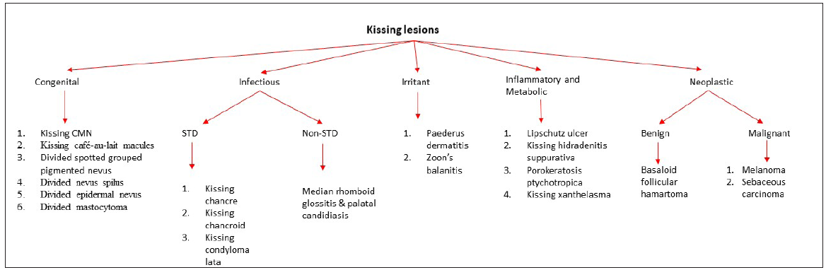
- Flowchart showing the classification of kissing lesions in dermatology. (CMN: Congenital melanocytic naevus, STD: Sexually transmitted disease.)
Conclusion
Kissing lesions are a rare morphology of dermatologic lesions with varied causes which must be kept in mind for an accurate diagnosis.
Declaration of patient consent
The authors certify that they have obtained all appropriate patient consent.
Financial support and sponsorship
Nil.
Conflicts of interest
There are no conflicts of interest.
Use of artificial intelligence (AI)-assisted technology for manuscript preparation
The authors confirm that there was no use of AI-assisted technology for assisting in the writing or editing of the manuscript and no images were manipulated using AI.
References
- Hidradenitis suppurativa presenting as genital “kissing lesions”. J Am Acad Dermatol. 2017;76:AB2.
- [Google Scholar]
- Flexural eczema versus atopic dermatitis. Dermatitis. 2015;26:109-15.
- [CrossRef] [PubMed] [Google Scholar]
- Kissing melanoma or kissing nevus of the penis? Urology. 2007;69(2):384.e5-7.
- [CrossRef] [PubMed] [Google Scholar]
- Kissing nevus of the penis: a case report and dermatoscopic findings. An Bras Dermatol. 2017;92:95-7.
- [CrossRef] [PubMed] [PubMed Central] [Google Scholar]
- Divided nevus of the eyelid: review of embryology, pathology and treatment. Am J Otolaryngol. 2013;34:223-9.
- [CrossRef] [PubMed] [Google Scholar]
- Kissing atypical melanocytic nevus of genital type of the labia majora in a young Bulgarian patient. What’s the best approach? Dermatol Rep. 2023;15:9667.
- [Google Scholar]
- Divided nevus of the eyelid: review of embryology, pathology and treatment. Am J Otolaryngol. 2013;34:223-9.
- [CrossRef] [PubMed] [Google Scholar]
- Divided nevus spilus and divided form of spotted grouped pigmented nevus. J Cutan Pathol.. 1979;6:507-12.
- [CrossRef] [PubMed] [Google Scholar]
- A case of epidermal nevi showing a divided form on the fingers. J Am Acad Dermatol.. 1993;29:281-2.
- [CrossRef] [PubMed] [Google Scholar]
- A case of solitary mastocytoma suggesting a divided form of mast cell nevus. J Dermatol.. 1989;16:402-4.
- [CrossRef] [PubMed] [Google Scholar]
- Congenital “kissing” lesions: nevus or “café au lait” spot? Revue de Stomatologie, de Chirurgie Maxillo-faciale et de Chirurgie Orale. 2016;117:433-7.
- [CrossRef] [PubMed] [Google Scholar]
- Hyperplastic candidosis on the palate developed as a ‘kissing’ lesion from median rhomboid glossitis. Braz J Otorhinolaryngol.. 2010;76:137.
- [CrossRef] [PubMed] [Google Scholar]
- Oral condyloma lata in secondary syphilis with kissing distribution. Actas Dermosifiliogr.. 2023;S0001-7310:00688-9.
- [Google Scholar]
- Kissing Lesions in Paederus Dermatitis. Am J Trop Med Hyg.. 2019;101:5.
- [CrossRef] [PubMed] [PubMed Central] [Google Scholar]
- Zoon balanitis: A comprehensive review. Indian J Sex Transm Dis AIDS.. 2016;37:129-38.
- [CrossRef] [PubMed] [PubMed Central] [Google Scholar]
- Ulcer of Lipschutz, a rare and unknown cause of genital ulceration. Facts Views Vis Obgyn.. 2018;10:55-7.
- [PubMed] [PubMed Central] [Google Scholar]
- Genitogluteal porokeratosis: a clinical review. Clin Cosmet Investig Dermatol.. 2018;11:219-29.
- [CrossRef] [PubMed] [PubMed Central] [Google Scholar]
- Unilateral xanthelasma forming a kissing-like lesion. J Dermatol.. 2005;32:220-2.
- [CrossRef] [PubMed] [Google Scholar]
- “Kissing” basaloid follicular hamartomas of the eyelid. J Cutan Pathol.. 2022;49:881-4.
- [CrossRef] [PubMed] [Google Scholar]
- Kissing tumors of sebaceous carcinoma on the eyelids in collision with melanocytic nevus: a case report. J Dermatol.. 2016;43:447-9.
- [CrossRef] [PubMed] [Google Scholar]


