Translate this page into:
Management of ear rim keloid with carbon dioxide laser
Correspondence Address:
D S Krupa Shankar
Department of Dermatology, Manipal Hospital, Airport Road, Bangalore - 560 017
India
| How to cite this article: Krupa Shankar D S, Gupta V. Management of ear rim keloid with carbon dioxide laser. Indian J Dermatol Venereol Leprol 2007;73:445 |
 |
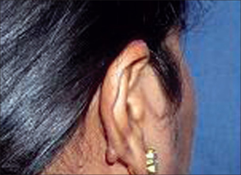
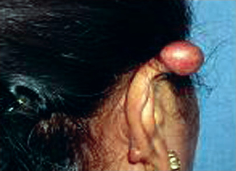
| Figure 2: Ear keloid after laser Surgery |
 |
| Figure 2: Ear keloid after laser Surgery |
 |
| Figure 1: Ear keloid before laser Surgery |
 |
| Figure 1: Ear keloid before laser Surgery |
Sir,
A 30 year-old housewife presented with itchy, painful swellings of eight months′ duration, on the ear lobe and posterior margin of the pinna. These swellings followed multiple ear piercings beginning from the lobe and extending along the rim of the ear 2-3 months after piercing.
The lesions had been treated with Cepain gel and clobetasol propionate ointment daily as well as with intralesional triamcinolone 10 mg/ml on two occasions a month apart, prior to consultation with no appreciable benefit. The patient is not a diabetic, hypertensive or atopic.
A diagnosis of the ear lobe and ear margin keloid was made. The nature of the condition was explained to the patient; topical, intralesional, cryotherapy, cold steel excision and laser excision were discussed and the pros and cons of each modality were weighed and explained. The patient made an informed decision in favor of laser excision [Figure - 1].
After aseptic precautions, the great auricular nerve was blocked. This was supplemented with sublesional lignocaine 2% in a manner that would expand the line of demarcation of the lesion. UML25 carbon dioxide laser was set in super-pulsed mode at 6.3 watts with a 0.1 mm handpiece. After running the laser incision around the base of the lesion, the keloid was grasped by tissue forceps and separated by a laser beam from its cutaneous anchorage [Figure - 2]. Oozing from the wound bed was controlled and defocused in continuous mode with the beam at 6 watts. The residual part of the keloidal tissue palpable in the wound floor was vaporized with the same super-pulsed settings described earlier.
The wound was dressed every 48 hours for one week followed by hydrocolloid dressings daily by the patient for three more weeks. The patient was followed up for a period of six months and no recurrences were noted at the operative site. The patient was advised against further ear piercing and to resort to clip-on ear jewellery.
Ear piercing is a long-standing tradition in many cultures. A fraction of those who pierce the ear develop infections, allergies to the inserted materials or keloids. Keloids result from the deposition of dense collagen bundles due to increased fibroblast activity. Up to 15-20% of all individuals are keloid-prone. Some races, notably Afro-Caribbean, are more keloid-prone than others.
Keloid excision followed by intralesional instillation of triamcinolone, bleomycin, [1] verapamil and 5-fluorouracil (5-FU) is one line of management. Intralesional cryotherapy alone and in combination with intralesional steroid therapy is well established. [2] Recurrences after surgery or intralesional therapy are common. [3] A variety of lasers have been used to treat keloids. [4],[5] Some authors have demonstrated encouraging results with CO 2 laser in keloids of the ear lobe while some others contest the same. CO 2 laser has some benefits over other modalities in terms of absence or delay in recurrence due to its positive effect on fibroblast secretion of growth factor. [4] Our patient is on follow-up and has been counselled to receive intralesional steroid therapy with cryotherapy at the earliest sign of recurrence.
| 1. |
Bodokh I, Brun P. The treatment of keloids with intralesional bleomycin. Ann Dermatol Venerol 1996;123:791-4
[Google Scholar]
|
| 2. |
Malakar S, Malakar R. Intralesional cryosurgery: Consequences, cautions and precautions. Indian J Dermatol 2000;45:100-1.
[Google Scholar]
|
| 3. |
Mutalik S. Treatment of keloids and hypertrophic scars. Indian J Dermatol Venereol Leprol 2005;71:3-8.
[Google Scholar]
|
| 4. |
Nowak KC, McCormack M, Koch RJ. The effect of superpulsed carbon dioxide laser energy on keloid and normal dermal fibroblast secretion of growth factors: A serum-free study. Plast Reconstr Surg 2000;105:2039-48.
[Google Scholar]
|
| 5. |
Cheng ET, Nowak KC, Koch RJ. Effect of blended carbon dioxide and erbium:YAG laser energy on preauricular and ear lobule keloid fibroblast secretion of growth factors: A serum-free study. Arch Facial Plast Surg 2001;3:252-7.
[Google Scholar]
|
Fulltext Views
2,457
PDF downloads
2,832


