Translate this page into:
Microcystic adnexal carcinoma
2 Department of Pathology, GGMC and Sir JJ Group of Hospitals, Mumbai, Maharashtra, India
Correspondence Address:
Veeral Manoj Aliporewala
Department of DVL, GGMC and Sir JJ Group of Hospitals, Mumbai, Maharashtra
India
| How to cite this article: Kura MM, Aliporewala VM, Bolde S, Bijwe S. Microcystic adnexal carcinoma. Indian J Dermatol Venereol Leprol 2020;86:595-596 |
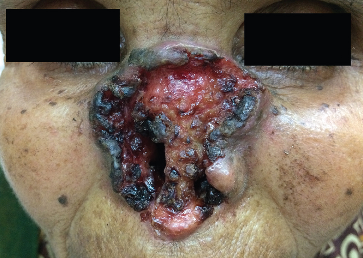
A 75-year-old female presented to our outpatient department with an ulcerative lesion on the face, gradually increasing for the last 4 years. Clinical examination revealed a large ulcer measuring ~ 6 × 8 cm, with raised, everted margins along with complete nasal destruction. The ulcer had involved the cheeks after eroding the entire nasal cartilage. [Figure - 1]. Histopathological examination demonstrated an uncircumscribed tumor with islands of basaloid cells in the upper dermis exhibiting follicular differentiation. The entire thickness of the lower dermis was filled with identical follicular basaloid cells along with numerous ill-formed ductal structures representing abortive ductal differentiation, suggestive of microcystic adnexal carcinoma.
 |
| Figure 1: Phagedenic ulcer causing destruction of the nose architecture |
Declaration of patient consent
The authors certify that they have obtained all appropriate patient consent forms. In the form, the patient has given her consent for her images and other clinical information to be reported in the journal. The patient understands that name and initials will not be published and due efforts will be made to conceal identity, but anonymity cannot be guaranteed.
Financial support and sponsorship
Nil.
Conflicts of interest
There are no conflicts of interest.
Fulltext Views
3,839
PDF downloads
2,086


