Translate this page into:
Moulages of J. J. Hospital
2 Department of Pathology, Sir J. J. Group of Hospitals, Mumbai, India
3 Department of Dermatology, Sir J. J. Group of Hospitals, Mumbai, India
Correspondence Address:
Rajiv Joshi
14 Jay Mahal, A Road, Churchgate, Mumbai - 400 020
India
| How to cite this article: Joshi R, D'Costa G, Kura MM. Moulages of J. J. Hospital. Indian J Dermatol Venereol Leprol 2010;76:583-588 |
Moulages of the Sir Jamshetjee Jeejeebhoy (Jj) Hospital, Mumbai
In a previous article [1] we described in brief the history of the dermatologic moulage, its origins from the early wax modeling known as ′Anatomica Plastica′ where minute details of the human anatomy were depicted by wax models to the next logical step of illustration of diseases by the use of wax models (Patho-anatomica plastica).
We described the origins of dermatologic moulage making in the early part of the 19 th century with pioneers like Martens of Jena, Germany, Elfinger from Vienna, and the Englishman Joseph Towne who in London made the moulages that form the subject for this paper. We traced the growth of the dermatologic moulage through the 19 th and early 20 th century as the preeminent teaching aid in dermato-venereology and its gradual decline after the Second World War.
Before we describe the moulages of the JJ Hospital, it is relevant here to give short biographical sketches of two individuals, both Englishmen, one the artist who created these moulages and the other, an equally famous artist and physician who we speculate might have been the driving force in getting these moulages to the JJ Hospital. The first is Joseph Towne of London and the second Henry Vandyke Carter, one time professor of Anatomy and Physiology and later principal of the Grant medical college and The Sir JJ Hospital, Bombay.
Joseph Towne: Wax Modeler Extraordinary of Guy′s Hospital, London [2],[3]
Joseph Towne (1806-79) was born in an artistic family and undoubtedly was strongly influenced by his father Thomas Towne, who, likely induced in his talented son a love for clay modeling. While still a teenager he built a 33 inch model of a skeleton that was anatomically accurate, having created the model over a period of about 2 years. This was submitted to the Society of Arts in London on the recommendation of the renowned surgeon from Guy′s hospital, Sir Astley Cooper (1768 - 1841) and won a silver medal.
Sir Astley Cooper was so impressed with the youngster′s ability for meticulous modeling that he recommended him to Benjamin Harrison who was the treasurer of the hospital, for appointment as medical illustrator and model maker. It was thus, that in February 1826, Towne was hired by Guy′s hospital to record rare and common medical conditions by drawings, models, and casts, a job that he was to hold for 53 years till his death in 1879.
In the five decades that he spent at Guy′s, he is known to have created at least 560 dermatological moulages, apart from numerous anatomical and other medical moulages. His models encompassed the entire spectrum of dermatological diseases and depicted the patient′s skin ailments in an exact, realistic, lifelike manner.
Making of moulages required close collaboration between physician and the artist and many of Towne′s moulages were based on patients of Thomas Addison (1793-1860). Addison established, at Guy′s hospital a centre renowned for dermatology and sent all interesting cases to Towne for having their models made. These superbly crafted dermatological moulages helped in no small way in establishing dermatology as a distinct discipline in the mid 19 th century.
Joseph Towne was an intensely secretive person and a recluse, and used to work alone locked in a room to which he held the only key. Although he did have a boy named Mills working for him as an errand boy and William Bouch as the model for his face casts, he never took on any apprentice nor taught his art to any one who could carry on after him and on his death in 1879, his techniques of moulaging died with him.
Towne had great loyalty to Guy′s hospital, his employer for over 50 years and refused to do work for any other hospitals in Britain. His work is therefore a single collection, namely the one at Guy′s hospital which is now housed in the Gordon Museum. He did however send work abroad, to India, Russia, Australia, and especially to the USA.
The collection of dermatological moulages of the JJ Hospital described in this article is most certainly part of the models that Towne made for overseas clients and were sent to Bombay, India.
Henry Vandyke Carter (1831-97)
Henry Vandyke Carter was born on 22 May 1831, the eldest son of the artist Henry Barlow Carter. After attending Hull Grammar School he came to London in 1847 to study medicine at St. George′s Hospital. He qualified M.R.C.S., L.S.A. in 1852.
In June 1853, he obtained a studentship in human and comparative anatomy at the Royal College of Surgeons. He was also, until July 1857, a demonstrator in Anatomy at St. George′s Hospital. The talent which he shared with his father and brother also opened up a career as an anatomical artist. He began his work for Henry Gray (1827-61), and in 1856, he commenced the illustrations for Henry Gray′s Anatomy (London, 1858).
In 1858 Carter joined the Bombay Medical Service and in May 1858 was gazetted professor of Anatomy and Physiology at Grant Medical College. He also served as assistant surgeon in the Jamshetjee Jeejeebhoy Hospital. From 1863 to 1872 he was Civil Surgeon at Satara. In 1872 he returned to Europe on furlough, studying leprosy in Norway. On his return to India in 1875, he was deputed to Kathiawar in Gujarat to investigate leprosy. In 1876 he took charge of the Goculdas Tejpal Hospital, Bombay (part of the Sir JJ Group of Hospitals). During the famine of 1877-78 he investigated a fever among the starving people, recognizing it as relapsing fever, and identifying, for the first time in India, the spirillum in the blood of its victims. In 1877, he was appointed as an acting principal (and later Principal) of Grant Medical College and Physician of the Jamshetjee Jeejeebhoy Hospital, Bombay.
H. V. Carter retired in 1888 and was subsequently appointed as the Honorary Deputy Surgeon-General and Honorary Surgeon to the Queen. He died at Scarborough North Yorkshire, UK on 4 May 1897.
H. V. Carter′s contributions to tropical pathology and dermatology included important studies of leprosy, mycetoma (which he recognised in 1860 as a fungal disease), and relapsing fever.
The Moulages of the Department of Dermatology at the Sir Jj Hospital, Mumbai
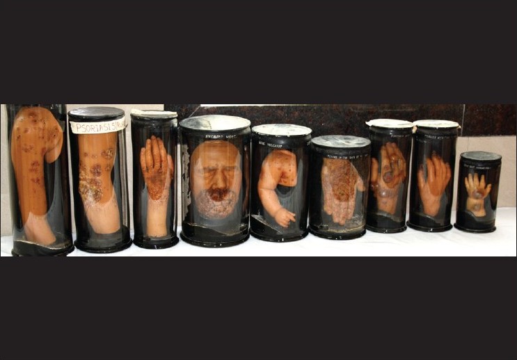
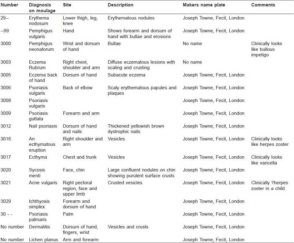
What follows is a description of the moulages of the department of Dermatology, Sir J. J. group of Hospitals, Mumbai) [Figure - 1] [Table - 1].
 |
| Figure 1 : Nine of the moulages showing the outer cylinder with dark backgrounds |

To the best of our knowledge this is the first report of a Moulage collection from India.
There are a total of 17 moulages in fairly well-preserved condition. These depict life like three-dimensional images of several skin diseases, namely psoriasis (5), eczema (3), herpes zoster/varicella (3), lichen planus (1), erythema nodosum (1), pemphigus vulgaris (1), pemphigus neonatorum (bullous impetigo) (1), sycosis barbae (1), and ichthyosis (1).
The moulages are enclosed in glass cylinders that are sealed at either ends with wooden boards to which the moulages are fixed. Half of the glass cylinder behind the moulage is painted black, while the other half facing the viewer is transparent for better view of the moulage.
The dimensions of the cylinders vary depending on the size of the moulage that it encloses. The smaller moulages (the dimensions are of the cylinder enclosing the moulage) are 30 cm in height by 15 cm in diameter (e.g. the moulage of palmar psoriasis) while the larger ones are 40 cm by 29 cm (eczema numbered 3003) and 42 cm by 17.5 cm (ichthyosis numbered 3029).
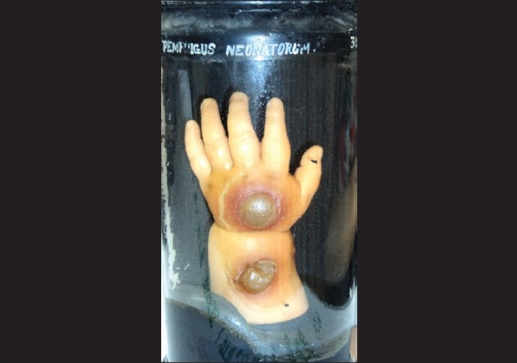
On the upper part of the cylinder above the moulage, on a black background is the diagnosis in white [Figure - 2].
 |
| Figure 2 : Moulage with diagnosis of pemphigus neonatorum (bullous impetigo), depicted on the upper part of the cylinder |
(In a few cases, the diagnosis written do not match the clinical appearance, e.g. moulage number 3016 is marked as ′an ecthymatous eruption′, but clinically is herpes zoster with typical vesicles; similarly, number 3017 too is marked as ′ecthyma′ but shows vesicles of varicella). It is most likely that the diagnosis was not written originally by the moulager but added on afterward by another person. (These diagnoses were most probably written in the pathology department under supervision of Dr. P. V. Gharpuray).
Also seen in white are numbers that identify the individual moulage. In this collection, 12 moulages show numbers that appear to be part of a series starting from 3000 to 3029. Three moulages show only part of the numbers, namely 29_ _, _ _ 98, and 30_ _. The remaining two moulages have no numbers.
At the base of the cylinder is a small metal tag on which is inscribed "Joseph Towne, Fecit, London." Fifteen of the moulages have this tag that marks them as made by Joseph Towne the famous English moulager of Guy′s hospital, London.
Fecit is Latin for "he made" or "made by" and refers to the engraver or artist who made the model.
The overall condition of the moulages is fairly good considering that they have been exposed to hot and humid conditions for all these years.
The color of most models has faded with yellowing of the wax; however, the lesions depicted are crisp and sharp [Figure - 3] and [Figure - 4].
 |
| Figure 3 : Moulage of acute vesicular dermatitis |
 |
| Figure 4 : Nail psoriasis with arthropathy |
One of the moulages (labeled ichthyosis and numbered 3029) shows degeneration of the wax with globs of whitish wax running over the surface and disintegration of the tip of the ring finger.
Moulages Housed in the Pathology Museum of the Sir Jj Hospital
An additional 61 moulages are housed in the pathology museum on the ground floor of the period building (built 1928-29) which houses the department of pathology of the Sir J. J. Hospital, Mumbai.
Most of these are in poor condition. Several have the diagnosis written on the top of the cylinder as well as the metal tag that identifies them as the work of Joseph Towne of London.
Three of these moulages will be discussed here as they are well preserved and depict vividly the disease or anatomical structure that they represent.
The first is marked 2999 and depicts beautifully swollen joints and deformed fingers of the hand in a case of rheumatoid arthritis.
The second marked 2987 shows TB dactylitis affecting the metacarpo-phalangeal joint of the thumb of the hand [Figure - 5].
 |
| Figure 5 : TB dactylitis |
The third moulage (3037) has the complete head with life-like depiction of hair on the scalp and beard areas.
Comments
The first chair of dermatology in India was established at the Sir J. J. Hospital, Bombay, in 1895 [4] and the first head of the Department of Dermatology was Major Cajetan Fernandez. This was the first department of dermatology in the whole of India until the second was established in 1923 at the School of Tropical Medicine in Calcutta, under the patronage of Col Acton and Dr. Ganapati Panja. [4]
Prior to this, dermatology in India did not exist as a distinct speciality as it did in Europe in the later half of the 19th century. Surgeons and physicians managed patients with skin diseases and at the Sir J. J. Hospital, Henry Vandyke Carter, had to his credit several publications in tropical medicine that included skin diseases like mycetoma and leprosy. Carter had a lasting interest in skin diseases and he was a medical illustrator himself, having done all the drawings for Henry Gray′s ′Anatomy′. Carter having worked in London in the 1850s must have been familiar with the dermatological work of Joseph Towne.
It is likely that the moulages of the JJ Hospital were ordered at the behest of Vandyke Carter who took over as the principal of the Grant Medical College and J. J. Hospital in 1877.
Joseph Towne died in 1879 and therefore the moulages must have been obtained before 1879, a period when Carter was in charge at the hospital.
Since 1952, department of dermatology at the JJ Hospital has been located in a separate building that housed the OPD on the ground floor and the wards for indoor patients on the first floor. The second floor had storage space for the department. This building is located far away from the main campus housing the hospital building and the buildings of other departments. Dr. H. J. Shroff, who joined as a house officer in dermatology in 1967 (went on to Head the department from 1982-97), recalls the presence of the moulages which were stored in glass cabinets on the first floor opposite the indoor wards, away from the daily hustle and bustle of the OPD. They remained there as mute spectators, were not used for teaching of dermatology to students or post-graduates, and were gradually forgotten by the succeeding generations of dermato-venereologists who passed through the department. It is likely that this isolation allowed the moulages to survive, or else they may have been destroyed to create space for a burgeoning department.
The skin OPD was shifted to the main OPD building of the hospital in 2005. It was during this shift from the old building that these long forgotten moulages resurfaced and were placed in glass cabinets in the new OPD. The first author recognized them for what they were and hence, he drafted this paper.
That these moulages are Towne′s moulages is clear as most of them carry the metal tag indicating so, however, what is not known is who ordered them and when did they come to India? It is likely that Henry Vandyke Carter may have played a role in getting them for the hospital; however, we have no records to prove that supposition.
The majority of the moulages repose in the pathology museum and it appears that the pathology department was the original recipient of the moulages. When and for what reason 17 of these moulages were transferred to the dermatology department is not known.
Dr. R. K. Gadgil, professor of pathology and head of department at the JJ Group of Hospital from 1958-73, recalls the presence of the moulages in the museum and affirms that his teacher professor P. V. Gharpuray (head of the department of pathology from 1927-51) was responsible for marking the moulages with a serial number and the diagnosis.
It is likely that these numbers were given as part of the inventory of the pathology museum which includes the pathology specimens in formalin and the wax models which repose side by side in the museum.
Conclusions
This paper describes a collection of 78 dermatological moulages made by Joseph Towne of London, that have survived and remained in the department of dermatology (17) and in the pathology museum (61) of the Sir JJ Hospital, Mumbai. The lifelike artistic and accurate rendering of skin diseases by these wax models is impressive and striking even after more than 130 years of their being cast.
This is the first description of a moulage collection from India but as Towne′s moulages are known to have been sent to other cities in India, namely Calcutta (now Kolkata) and Madras (now Chennai) it is possible that collections of moulages survive in these cities in the old Medical College museums and it is a hope that this article may lead to the cataloging and descriptions of these long forgotten historical treasures of dermatology!
| 1. |
Joshi R. Moulages in dermatology and venereology. Indian J Dermatol Venereol Leprol 2010;76:434-8.
[Google Scholar]
|
| 2. |
Atherton DJ. Joseph Towne: wax modeler extraordinary: J Am Acad Dermatol 1980;3:311-6.
[Google Scholar]
|
| 3. |
Alberti Samuel AJ. Wax bodies: art and anatomy in victorian medical museums. Mus Hist J 2009;2:7-36.
[Google Scholar]
|
| 4. |
Thappa DM. History of dermatology, venereology and leprology in India. J Postgrad Med 2002;48:160-5.
[Google Scholar]
|
Fulltext Views
3,814
PDF downloads
3,187


