Translate this page into:
Multiple annular erythematous plaques on the back
2 Department of Dermatology, National Taiwan University Hospital, Taiwan, China
Correspondence Address:
Wang-Cheng Ko
Department of Dermatology, Show-Chwan Memorial Hospital, No.542, Sec, 1 Chung-Shang Rd. Changhua, Taiwan
China
| How to cite this article: Chou WT, Tsai TF, Hung CM, Ko WC. Multiple annular erythematous plaques on the back. Indian J Dermatol Venereol Leprol 2011;77:727-728 |
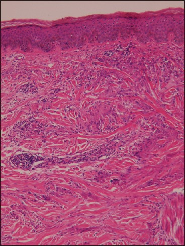
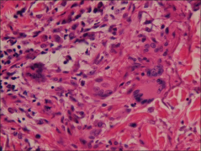
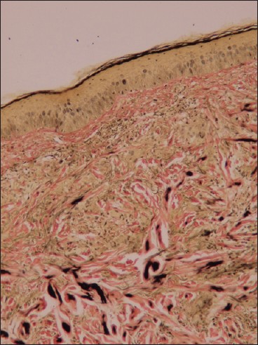
A 66-year-old man was referred to our clinic with multiple annular plaques on his back for 3 months. Initially, he found several asymmetric pruritic papules over his back and these papules spread gradually and coalesced to form large irregular plaques. The center of the lesion showed mild atrophy with peripheral elevated borders [Figure - 1]. Incision biopsy was performed on the elevated margin of the plaque and the sample was sent for histological examination. Microscopic examination showed basal hyper-pigmentations and granulomatous infiltrates of multinucleated giant cells in the upper and mid dermis [Figure - 2] and [Figure - 3]. Neither solar elastosis nor necrobiosis was found and there is no mucin deposit in the dermis. Besides, perivascular granulomatous changes were seen without vasculitis. Elastic-van Gieson′s stain showed decreasing elastic fiber densities in the mid-dermis and fragmentation of elastic fibers with elastophagocytosis [Figure - 4].
 |
| Figure 1: There were multiple annular erythematous plaques with elevated borders and confluent tendency on the back. The center of the lesion showed mild atrophy |
 |
| Figure 2: Basal hyperpigmentations and granulomatous infiltrates were presented in the upper and mid-dermis. No vasculitis was seen in the dermis. (H and E, ×100) |
 |
| Figure 3: Several multinucleated giant cells with elastophagocytosis are distributed in the mid-dermis. (H and E, ×400) |
 |
| Figure 4: Elastic van Gieson's stain showed decreasing elastic fiber densities in the mid-dermis and fragmentation of elastic fibers with elastophagocytosis. (Elastic van Gieson's stain, ×100) |
On review of the system, the patient was found to have type 2 diabetic mellitus under regular treatment for last 10 years. He was a farmer and had long-term sun exposure in the past 40 years.
What is Your Diagnosis?
| 1. |
Hanke CW, Bailin PL, Roenigk HH Jr. Annular elastolytic giant cell granuloma: A clinicopathologic study of five cases and a review of similar entities. J Am Acad Dermatol 1979;1:413-21.
[Google Scholar]
|
| 2. |
Klemke CD, Siebold D, Dippel E. Generalised annular elastolytic giant cell granuloma. Dermatology 2003;207:420-2.
[Google Scholar]
|
| 3. |
Limas C. The spectrum of primary cutaneous elastolytic granulomas and their distinction from granuloma annulare: A clinicopathological analysis. Histopathology 2004;44:277-82.
[Google Scholar]
|
| 4. |
Müller FB, Groth W. Annular elastolytic giant cell granuloma: A prodromal stage of mid-dermal elastolysis? Br J Dermatol 2007;156:1377-9.
[Google Scholar]
|
| 5. |
Boussault P, Tucker ML, Weschler J. Primary cutaneous CD4+ small/medium-sized pleomorphic T-cell lymphoma associated with an annular elastolytic giant cell granuloma. Br J Dermatol 2009;160:1126-8.
[Google Scholar]
|
Fulltext Views
2,366
PDF downloads
1,483


