Translate this page into:
Multiple floating fat balls on the right lower leg
Correspondence Address:
Shin Yi-Chin
Department of Dermatology, Chang Gung Memorial Hospital, 199, Tun-Hwa North Road, Taipei 105, Taiwan
China
| How to cite this article: Yi-Hsin H, Ya-Wen H, Yi-Chin S. Multiple floating fat balls on the right lower leg. Indian J Dermatol Venereol Leprol 2011;77:731 |
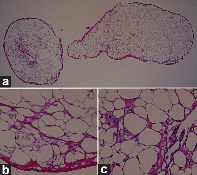
A 48-year-old woman presented with multiple mobile subcutaneous nodules on the right lower extremity for 1 year. The first subcutaneous nodule developed on the right ankle, increased in number, and spread to the proximal part with linear arrangement. The nodules could be pushed and displaced with greatest mobility up to 2-3 cm in the vertical direction. These lesions were asymptomatic except for mild tenderness on palpation. No worthy trauma history was mentioned. A clinical examination revealed multiple highly migrating rubbery mobile subcutaneous nodules [Figure - 1]a. Other physical, ophthalmological, and neurological examinations were all normal. Laboratory tests, including hemogram, biochemistry data, and autoimmune screening (antinuclear antibody, anti-Scl70 antibody), were all within normal limits. Computed tomography of the right lower leg revealed multiple subcutaneous calcifications [Figure - 1]b. Two adjacent lesions were taken from the right lower leg for histological examination. Gross examination revealed two bean-size orange-yellow spheral-shaped adipose nodules with firm consistency [Figure - 1]c. The specimen underwent histological examination with hematoxylin-eosin [Figure - 2].
 |
| Figure 1: (a) Multiple mobile subcutaneous nodules on the right lower leg. (b) Computed tomography of the right lower leg showed multiple subcutaneous calcifications. (c) Gross examination showed two bean-sizes orange-yellow spheral-shaped adipose nodules |
 |
| Figure 2: Skin biopsy of the specimen showing (a) two well-defined, oval- shaped fat tissue surrounded by condensed fibrous tissue (H and E, ×20). (b) Peripherally, focal lipomembrance change was noted (H and E, ×400). (c) Fat necrosis with calcification and chronic inflammation was evident in the central part of the nodules (H and E, ×400) |
What is Your Diagnosis?
| 1. |
Toshiyuki Y, Kiyoshi N, Miho T. Nodular cystic fat necrosis with systemic sclerosis. Eur J Dermatol 2004;14:353-5.
[Google Scholar]
|
| 2. |
Felipo F, Vaquero M, Del Agua C. Pseudotumoral encapsulated fat necrosis with diffuse pseudomembranous degeneration. J Cutan Pathol 2004;31:565-7.
[Google Scholar]
|
| 3. |
Hurt MA, Santa-Cruz DJ. Nodular-cystic fat necrosis: A reevaluation of the so-called mobile encapsulated lipoma. J Am Acad Dermatol 1989;21:493-8.
[Google Scholar]
|
| 4. |
Rahsan G, Ulku K. An unusual appearance of traumatic fat necrosis: Floating fat balls. Eur J Radiol 2008;66:43-5.
[Google Scholar]
|
| 5. |
Hendrick SJ, Silverman AK, Solomon AR, Headington JT. Alpha 1-antitrypsin deficiency associated with panniculitis. J Am Acad Dermatol 1988;18:684-92.
[Google Scholar]
|
Fulltext Views
5,634
PDF downloads
3,510


