Generic selectors
Exact matches only
Search in title
Search in content
Post Type Selectors
Search in posts
Search in pages
Filter by Categories
15th National Conference of the IAOMFP, Chennai, 2006
Abstract
Abstracts from current literature
Acne in India: Guidelines for management - IAA Consensus Document
Addendum
Announcement
Art & Psychiatry
Article
Articles
Association Activities
Association Notes
Award Article
Book Review
Brief Report
Case Analysis
Case Letter
Case Letters
Case Notes
Case Report
Case Reports
Clinical and Laboratory Investigations
Clinical Article
Clinical Studies
Clinical Study
Commentary
Conference Oration
Conference Summary
Continuing Medical Education
Correspondence
Corrigendum
Cosmetic Dermatology
Cosmetology
Current Best Evidence
Current Issue
Current View
Derma Quest
Dermato Surgery
Dermatopathology
Dermatosurgery Specials
Dispensing Pearl
Do you know?
Drug Dialogues
e-IJDVL
Editor Speaks
Editorial
Editorial Remarks
Editorial Report
Editorial Report - 2007
Editorial report for 2004-2005
Errata
Erratum
Focus
Fourth All India Conference Programme
From Our Book Shelf
From the Desk of Chief Editor
General
Get Set for Net
Get set for the net
Guest Article
Guest Editorial
History
How I Manage?
IADVL Announcement
IADVL Announcements
IJDVL Awards
IJDVL AWARDS 2015
IJDVL Awards 2018
IJDVL Awards 2019
IJDVL Awards 2020
IJDVL International Awards 2018
Images in Clinical Practice
Images in Dermatology
In Memorium
Inaugural Address
Index
Knowledge From World Contemporaries
Leprosy Section
Letter in Response to Previous Publication
Letter to Editor
Letter to the Editor
Letter to the Editor - Case Letter
Letter to the Editor - Letter in Response to Published Article
LETTER TO THE EDITOR - LETTERS IN RESPONSE TO PUBLISHED ARTICLES
Letter to the Editor - Observation Letter
Letter to the Editor - Study Letter
Letter to the Editor - Therapy Letter
Letter to the Editor: Articles in Response to Previously Published Articles
Letters in Response to Previous Publication
Letters to the Editor
Letters to the Editor - Letter in Response to Previously Published Articles
Letters to the Editor: Case Letters
Letters to the Editor: Letters in Response to Previously Published Articles
Media and news
Medicolegal Window
Messages
Miscellaneous Letter
Musings
Net Case
Net case report
Net Image
Net Images
Net Letter
Net Quiz
Net Study
New Preparations
News
News & Views
Obituary
Observation Letter
Observation Letters
Oration
Original Article
ORIGINAL CONTRIBUTION
Original Contributions
Pattern of Skin Diseases
Pearls
Pediatric Dermatology
Pediatric Rounds
Perspective
Presedential Address
Presidential Address
Presidents Remarks
Quiz
Recommendations
Regret
Report
Report of chief editor
Report of Hon : Treasurer IADVL
Report of Hon. General Secretary IADVL
Research Methdology
Research Methodology
Resident page
Resident's Page
Resident’s Page
Residents' Corner
Residents' Corner
Residents' Page
Retraction
Review
Review Article
Review Articles
Reviewers 2022
Revision Corner
Self Assessment Programme
SEMINAR
Seminar: Chronic Arsenicosis in India
Seminar: HIV Infection
Short Communication
Short Communications
Short Report
Snippets
Special Article
Specialty Interface
Studies
Study Letter
Study Letters
Supplement-Photoprotection
Supplement-Psoriasis
Symposium - Contact Dermatitis
Symposium - Lasers
Symposium - Pediatric Dermatoses
Symposium - Psoriasis
Symposium - Vesicobullous Disorders
SYMPOSIUM - VITILIGO
Symposium Aesthetic Surgery
Symposium Dermatopathology
Symposium-Hair Disorders
Symposium-Nails Part I
Symposium-Nails-Part II
Systematic Review and Meta-Analysis
Systematic Reviews and Meta-analyses
Systematic Reviews and Meta-analysis
Tables
Technology
Therapeutic Guideline-IADVL
Therapeutic Guidelines
Therapeutic Guidelines - IADVL
Therapeutics
Therapy
Therapy Letter
Therapy Letters
View Point
Viewpoint
What’s new in Dermatology
View/Download PDF

Translate this page into:

Quiz
ARTICLE IN PRESS
doi:
10.25259/IJDVL_939_2023

Multiple hairy nodules in an adolescent girl

Department of Dermatology and Venereology and Pathology, All India Institute of Medical Sciences, Bhubaneswar, Odisha, India
Department of Pathology, All India Institute of Medical Sciences, Bhubaneswar, Odisha, India

Corresponding author: Dr. Priyansh Gupta, Department of Dermatology and Venereology, All India Institute of Medical Sciences, Bhubaneswar, Odisha, India. priyansh.gupta2412@gmail.com

Licence
This is an open-access article distributed under the terms of the Creative Commons Attribution-Non Commercial-Share Alike 4.0 License, which allows others to remix, transform, and build upon the work non-commercially, as long as the author is credited and the new creations are licensed under the identical terms.

How to cite this article: Gupta P, Sirka CS, Shetty M, Rout AN. Multiple hairy nodules in an adolescent girl. Indian J Dermatol Venereol Leprol. doi: 10.25259/IJDVL_939_2023

A thirteen-year-old girl, with a known case of ventricular septal defect, presented with a two-year duration of multiple skin-coloured to hyperpigmented nodules over her face, trunk and extremities. The lesions were asymptomatic, without any surface erosion or ulceration. On examination, multiple (eleven) firm-to-hard tender nodules of size ranging from 0.5 to 1.5 cm were present over the sites mentioned above. In addition, the skin-overlying nodules were pigmented and associated with hypertrichosis [Figures 1a and 1b]. The nodules on the face and thigh were bony hard, suggestive of calcification, which was later confirmed on X-ray evaluation of the skull and thigh [Figure 2]. Examination of the oral cavity showed a solitary, small, whitish, non-tender nodule on the left lateral aspect of the tongue [Figure 3]. There was no lymphadenopathy, and the rest of the mucocutaneous examination revealed no abnormality. Dermoscopy of the cutaneous nodules showed an increased pigment network with areas of perifollicular sparing [Figure 4]. An incisional biopsy was done from the nodule present over the left thigh for histopathological examination.
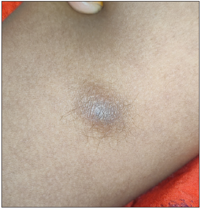
Ill-defined hyperpigmented subcutaneous nodule over left thigh with overlying hypertrichosis.
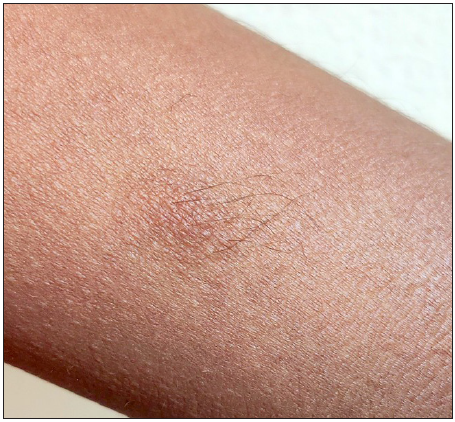
Figure 1a:
Ill-defined hyperpigmented subcutaneous nodule over left thigh with overlying hypertrichosis.
Nodular lesion present over the left arm with overlying hyperpigmentation and hypertrichosis.
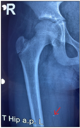
Figure 1b:
Nodular lesion present over the left arm with overlying hyperpigmentation and hypertrichosis.
X-ray of the right thigh showing radio-opaque shadow (red arrow) suggestive of calcification.
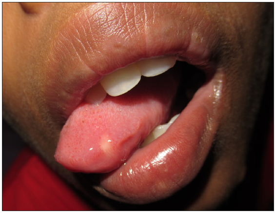
Figure 2:
X-ray of the right thigh showing radio-opaque shadow (red arrow) suggestive of calcification.
Oral cavity showing a solitary white nodule present on the lateral aspect of the tongue.
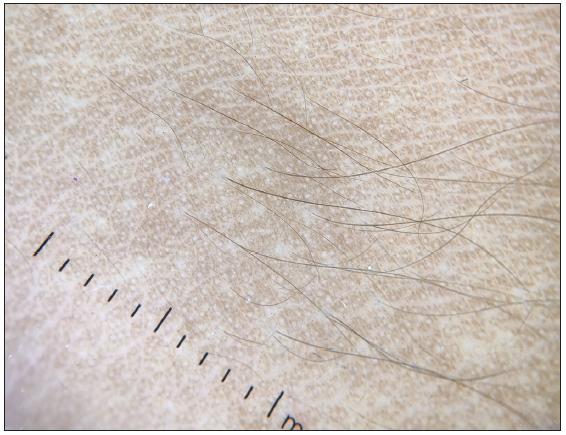
Figure 3a:
Oral cavity showing a solitary white nodule present on the lateral aspect of the tongue.
Dermoscopic examination showed diffuse structureless pigmentation with areas of perifollicular sparing (DermLite DL4 dermoscopy 10x).
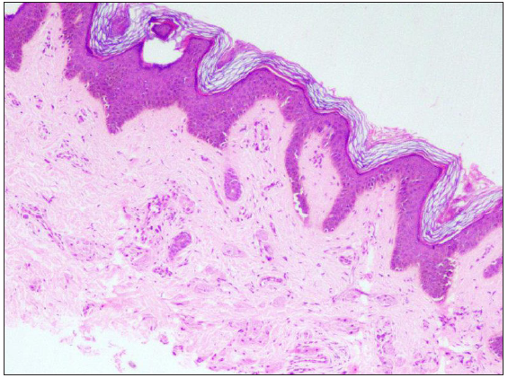
Figure 3b:
Dermoscopic examination showed diffuse structureless pigmentation with areas of perifollicular sparing (DermLite DL4 dermoscopy 10x).
Histopathological examination showed normal epidermis and scattered large cells in the dermis (Haematoxylin & Eosin, 100x).
Figure 4a:
Histopathological examination showed normal epidermis and scattered large cells in the dermis (Haematoxylin & Eosin, 100x).
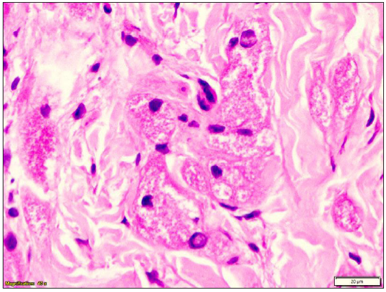
On higher magnification, large polygonal cells with eosinophilic granular cytoplasm and central nuclei in the upper dermis suggest granular cell tumour (Haematoxylin & Eosin, 400x).
Figure 4b:
On higher magnification, large polygonal cells with eosinophilic granular cytoplasm and central nuclei in the upper dermis suggest granular cell tumour (Haematoxylin & Eosin, 400x).

Histopathological Examination

The epidermis was unremarkable. In the dermis, scattered, large, round to polygonal cells with eosinophilic granular cytoplasm and central nuclei were present [Figures 4a and 4b]. The infiltrate was limited to the upper dermis without any subcutaneous tissue involvement. Immunohistochemistry was positive for CD68 [Figure 5] and S100 and negative for CD1a [Figure 4].
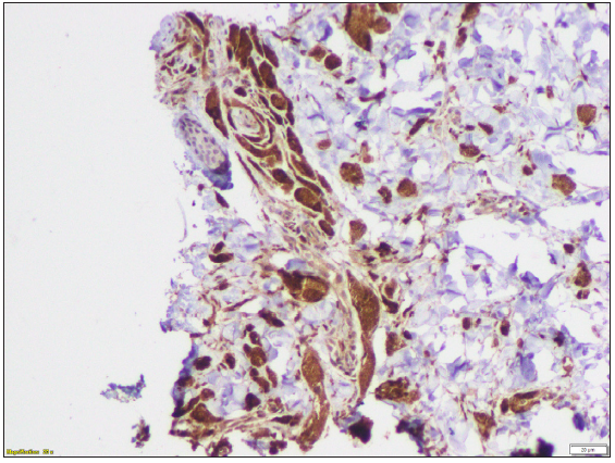
Immunohistochemistry of the histopathology showing tumour cells with CD68 positivity (Immunohistochemistry, 200x).
Figure 5:
Immunohistochemistry of the histopathology showing tumour cells with CD68 positivity (Immunohistochemistry, 200x).

Question

What is your diagnosis?

Answer

Diagnosis: Hairy granular cell tumour

Discussion

Granular cell tumour (GCT) is a rare benign neoplasm originating from Schwann cells. There is a female preponderance and it commonly affects middle-aged individuals.1 The most accepted pathogenesis is the altered metabolism of Schwann cells, but origin from histiocytes, fibroblasts, or myoepithelial cells has also been postulated.2 It manifests as a solitary subcutaneous or submucosal nodule commonly involving the tongue or internal organs. Around one-fifth of the cases present with multiple lesions.2 Pointers towards diagnosing GCT in our case include multiple lesions, overlying hypertrichosis and mucosal lesions. The other differentials to be considered in lesions with overlying hypertrichosis include plexiform neurofibroma, leiomyoma, melanocytic nevus, smooth muscle hamartoma and eccrine angiomatous hamartoma. The nodules of leiomyoma and neurofibroma are firm in consistency and spare mucosa, while melanocytic nevus and smooth muscle hamartoma lack isolated nodules as in our case. Eccrine angiomatous hamartoma can have hyperhidrosis in addition to hypertrichosis and shows hyperplastic eccrine glands on histopathology. Moreover, hypertrichosis on cutaneous lesions of GCTs is extremely rare, with only one case reported.3 Increased stimulation of hair follicles and prolongation of the anagen growth cycle can be the possible mechanism. However, the exact mechanism needs to be established. Another interesting finding in our case was the calcification of the lesions over the face and thighs. Previous literature reported calcification in colonic tumours as well as seller Granular cell tumour (GCT).4,5 However, there are no reports of calcified cutaneous granular cell tumours to date.

This case highlights the importance of keeping GCT as a differential when one comes across hairy nodules on the skin, especially if mucosa is involved. Previous literature mentioned the association of GCT with Noonan syndrome, neurofibromatosis type 1 and LEOPARD syndrome.6 Our patient also had a ventricular septal defect without any other features of LEOPARD syndrome. The common origin of cardiac septum and schwann cells from neural crest cells might explain the association but needs further validation.7 Nevertheless, it is prudent to know about associated syndromes so that proper evaluation can be done, which can prevent major cardiac events in future. The GCTs are mostly benign and rarely show malignant behaviour.2 The prognosis was explained to the girl’s parents, and she is under regular follow-up.

Declaration of patient consent

The authors certify that they have obtained all appropriate patient consent.

Financial support and sponsorship

Nil.

Conflicts of interest

There are no conflicts of interest.

Use of artificial intelligence (AI)-assisted technology for manuscript preparation

The authors confirm that there was no use of artificial intelligence (AI)-assisted technology for assisting in the writing or editing of the manuscript and no images were manipulated using AI.

References

  1. , , , . Granular cell tumour of the tongue in a 14-year-old boy: Case report. Acta Otorhinolaryngologica Italica. 2011;31:186-9.
    [PubMed] [PubMed Central] [Google Scholar]
  2. , , , , , . Granular cell tumour a study of 42 cases and systemic review of the literature. Pathol Res Pract. 2020;216:152865.
    [CrossRef] [PubMed] [Google Scholar]
  3. , , . “Hairy” granular cell tumour. Pediatr Dermatol. 2010;27:545-6.
    [CrossRef] [PubMed] [Google Scholar]
  4. , . Granular cell tumour of the cecum with extensive hyalinization and calcification: A case report. World J Gastroenterol. 2009;15:3315-8.
    [CrossRef] [PubMed] [PubMed Central] [Google Scholar]
  5. , , , , , . A case of symptomatic granular cell tumour of the sellar region with large calcification. Neuropathology. 2017;37:371-4.
    [CrossRef] [PubMed] [Google Scholar]
  6. , , . Granular Cell Tumour. Indian J Otolaryngol. 2022;34:69-70.
    [Google Scholar]
  7. , , , , , . Oral granular cell tumour: A multicentric study of 56 cases and a systematic review. Oral Dis. 2020;26:573-89.
    [CrossRef] [PubMed] [Google Scholar]
Show Sections