Translate this page into:
Pacinian neurofibroma: A rare neurogenic tumor
2 Department of Pathology, Jawaharlal Institute of Postgraduate Medical Education and Research (JIPMER), Pondicherry - 605 006, India
Correspondence Address:
Devinder Mohan Thappa
Department of Dermatology and STD, JIPMER, Pondicherry - 605 006
India
| How to cite this article: Nath AK, Timshina DK, Thappa DM, Basu D. Pacinian neurofibroma: A rare neurogenic tumor. Indian J Dermatol Venereol Leprol 2011;77:204-205 |
Sir,
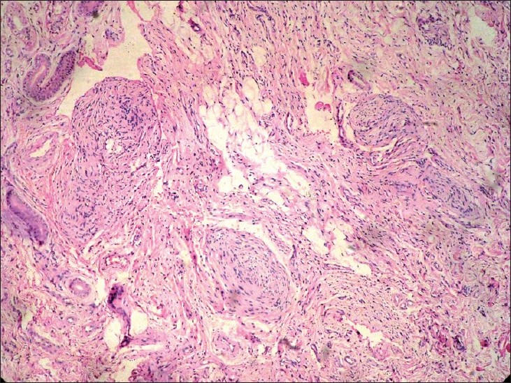
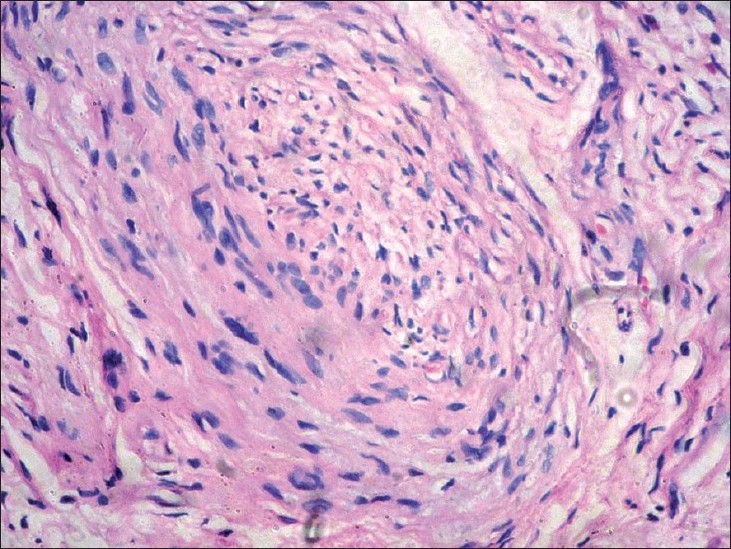
Pacinian neurofibroma (PN) is a rare tumor of neural origin histopathologically characterized by the formation of components resembling Pacinian corpuscles within the lobules of the tumor. [1] It usually occurs as a solitary nodule, most often on the hands and feet, where Pacinian corpuscles are usually concentrated. [2],[3] Lesions may occur elsewhere also. [1],[2] Diagnosis is established by histopathology features, which may be variable. We report a case of PN which presented as a slowly growing tumor on the forearms.A 17-year-old boy presented with an asymptomatic, well-defined, irregular, sessile, slow-growing tumor [Figure - 1] on the extensor aspect of right forearm of 3 years duration. The onset was spontaneous without any history of antecedent trauma. The swelling was fully excised earlier at a local hospital, but it recurred and kept on increasing in size. There were hyperpigmentation and growth of coarse, terminal hair on the surface of the swelling. On palpation, the tumor was non-tender, firm, lobulated, and freely mobile with negative "button holing" sign. Histopathological examination of an incisional biopsy specimen revealed non-encapsulated but well-delineated dermal lobules composed of relatively hypocellular central core surrounded by multiple (up to 20) layers of collagenous lamellae [Figure - 2]. The lobules contained elliptical or spindle-shaped nuclei both in the central core and in the surrounding lamellae [Figure - 3]. There was merging of the concentric lamellae with the collagen fibers of the adjacent dermis. The tissue around the lobules was cellular and contained poorly formed nerve bundles. There were normal eccrine glands in some areas, but no vascular spaces were seen. Minimal mucinous alteration of the stroma was seen. S100 staining was moderately reactive, which confirmed the neural origin of the tumor. CD34 staining was done and it was negative. A diagnosis of PN was made based upon the above-mentioned features. The patient was referred to the plastic surgery department for excision of the tumor.
 |
| Figure 1: Well-defined, irregular, sessile tumor on the extensor aspect of right forearm |
 |
| Figure 2: Photomicrograph showing well-delineated dermal lobules composed of relatively hypocellular central core surrounded by multiple (up to 20) layers of collagenous lamellae. Elliptical or spindle-shaped nuclei are seen both in the central core and in the surrounding lamellae (H and E, ×100) |
 |
| Figure 3: Close up image of Pacinian corpuscle like formations (H and E, ×200) |
PN was first described by Thoma in 1894, later by Prichard and Custer in 1952 as well as by Prose et al, in 1957. [3] Over the years, many authors have either described other tumors as PN or described PN under various other terms, some of which are used synonymously while the others are misnomer or represent other tumors. Fibrous (dermal) perineurioma (perineurioma refers to tumors of perineural cells) and sclerosing (subcutaneous) perineurioma have been recently used to describe PN; however, some authors feel that the term pacinian perineurial cell fibroma would better represent this tumor. [4] The term pacinioma represents hamartomatous overgrowth of mature Vater-Pacinian corpuscles. [5] PN should be differentiated from Pacinian hypertrophy and Pacinian hyperplasia where the classic structure of Pacinian corpuscles is well maintained. PN, on the other hand, exhibits Pacinian corpuscles-like differentiation within a myxoid stroma. [6],[7] PN is different from ordinary neurofibroma. Ordinary neurfibroma may show occasional mature Pacinian corpuscles, but typical histopathology of PN is not seen in ordinary neurofibroma. [6]
PN usually presents as solitary, firm, well-marginated, mobile nodule. [1],[2] Multiple lesions are rare. [1] Other morphological pattern that have been described in PN are papular lesions, ulcerated tumor, pedunculated nodule, multiple soft plaques, or just surface pigmentation. Most often, they occur on the hands and feet, but other reported sites of occurrence are buttocks, neck, flank, maxilla, arm, cheek, and sacrococcygeal region. [1],[2] Pressure on the nerve bundles may cause pain. [3] Multiple PNs have been associated with marked vascular changes of the glomus type of arteriovenous anastomosis. [2] No association with neurofibromatosis has been found. [1]
Histopathologically, a typical PN tumor is well demarcated, often an encapsulated mass containing round or ovoid lobules, each showing a central homogeneous, acellular, eosinophilic core surrounded by as many as 30 pale-staining, concentric collagenous lamellae. [3] These formations greatly resemble Pacinian corpuscles. [8] The size of these corpuscles is variable. The superficial corpuscles tend to be smaller (4-7 lamellae). [3] The more immature one′s have many cellular elements with spindle-shaped nuclei. [2] However, the more mature PN shows histopathology like mature Pacinian corpuscles. [2] There may be merging of the concentric lamellae with the collagen fibers of adjacent dermis. [3] In some tumors, the tissue around the lobules is cellular and contains poorly formed nerve bundles. [8] In such areas, the lobules contain numerous, elliptic or spindle-shaped nuclei both in the central core and in the surrounding lamellae; nuclei are reduced in number in mature lesions. Mucinous alteration of the stroma may be seen. [8]
PN is a very rare tumor. To the best of our knowledge, only single case of PN of the scalp has been reported from India. [9] Thus our case is the second report of this rare tumor from India.
| 1. |
McCormack K, Kaplan D, Murray JC, Fetter BF. Multiple hairy pacinian neurofibromas (nerve-sheath myxomas). J Am Acad Dermatol 1988;18:416-9.
[Google Scholar]
|
| 2. |
Levi L, Curri SB. Multiple Pacinian neurofibroma and relationship with the finger-tip arterio-venous anastomoses. Br J Dermatol 1980;102:345-9.
[Google Scholar]
|
| 3. |
Bennin B, Barsky S, Salgia K. Pacinian neurofibroma. Arch Dermatol 1976;112:1558.
[Google Scholar]
|
| 4. |
Reed RJ, Pulitzer DR. Tumors of neural tissue. In: Elder DE, editor. Lever's Histopathology of the Skin. 10 th ed. New Delhi: Wolters Kluwer/Lippincott Williams and Wilkins; 2009. p. 1133-34.
th ed. New Delhi: Wolters Kluwer/Lippincott Williams and Wilkins; 2009. p. 1133-34.'>[Google Scholar]
|
| 5. |
Weedon D, Strutton G, editors. Skin Pathology. 2 nd ed. China: Churchill/Livingstone Elsevier Science Ltd; 2002. p. 984.
[Google Scholar]
|
| 6. |
Fraitag S, Gherardi R, Wechsler J. Hyperplastic pacinian corpuscles: An uncommonly encountered lesion of the hand. J Cutan Pathol 1994;21:457-60.
[Google Scholar]
|
| 7. |
Yan S, Horangic NJ, Harris BT. Hypertrophy of pacinian corpuscles in a young patient with neurofibromatosis. Am J Dermatopathol 2006;28:202-4.
[Google Scholar]
|
| 8. |
Lever WF, Schaumberg-Lever G. Histopathology of the Skin. 6 th ed. Philadelphia: J.B. Lippincott Company; 1983. p. 673-4.
[Google Scholar]
|
| 9. |
Deshpande GU, Bhatoe HS, Rai R, Panicker NK, Ramji R. Pacinian neurofibroma of scalp. Med J Armed Forces India 1997;53:135-6.
[Google Scholar]
|
Fulltext Views
4,477
PDF downloads
3,187


