Translate this page into:
Painful tumors of the skin – from ENGLAND to LEND AN EGG to BLEND TAN EGG
Correspondence Address:
M Ramesh Bhat
Department of Dermatology, Father Muller Medical College, Mangalore - 575 002, Karnataka
India
| How to cite this article: Bhat M R, George AA, Jayaraman J. Painful tumors of the skin – from ENGLAND to LEND AN EGG to BLEND TAN EGG. Indian J Dermatol Venereol Leprol 2019;85:231-234 |
Acronyms
The acronyms for painful tumors of the skin always evoke an interest among students and teachers of dermatology. In fact, the main author of this article was asked during his Post Graduate examination about the acronym ENGLAND. Over the years with the addition of newer entities, new acronyms have been developed. The only common symptom attributed to these heterogeneous benign tumors is pain. We also had the opportunity to publish a few case reports related to atypical presentations of these tumors.[1],[2],[3] The acronym ENGLAND or GLENDA is often used to recall these tumors – eccrine spiradenoma, neuroma, glomus tumor, leiomyoma, angiolipoma, neurilemmoma and dermatofibroma. Naversen et al. modified the acronym “LEND AN EGG” with the addition of endometrioma and granular cell tumor.[4] With the addition of blue rubber bleb nevus and tufted angioma to this list, presently the acronym stands as “BLEND TAN EGG.”[2] In this article, we have briefly described the clinical features and analyzed the pathogenesis of pain in these tumors [Table - 1].
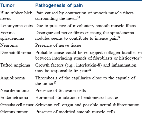
Blue Rubber Bleb Nevus
It is characterized by cutaneous and gastrointestinal venous malformations. Skin lesions have a cyanotic bluish appearance with soft elevated nipple-like projections. Nocturnal pain is characteristic.[5]
Leiomyoma
This tumor develops from smooth muscles.[6] Solitary and multiple piloleiomyomas are derived from arrector pili muscle and angioleiomyomas are derived from muscles of vein.[7] These lesions present as solitary or grouped red, pink, purple, brown, waxy or translucent nodules [Figure - 1] and [Figure - 2]. On histopathological examination, interlacing bundles of smooth muscle bundles with characteristic eel-shaped nuclei are seen [Figure - 3].[4]
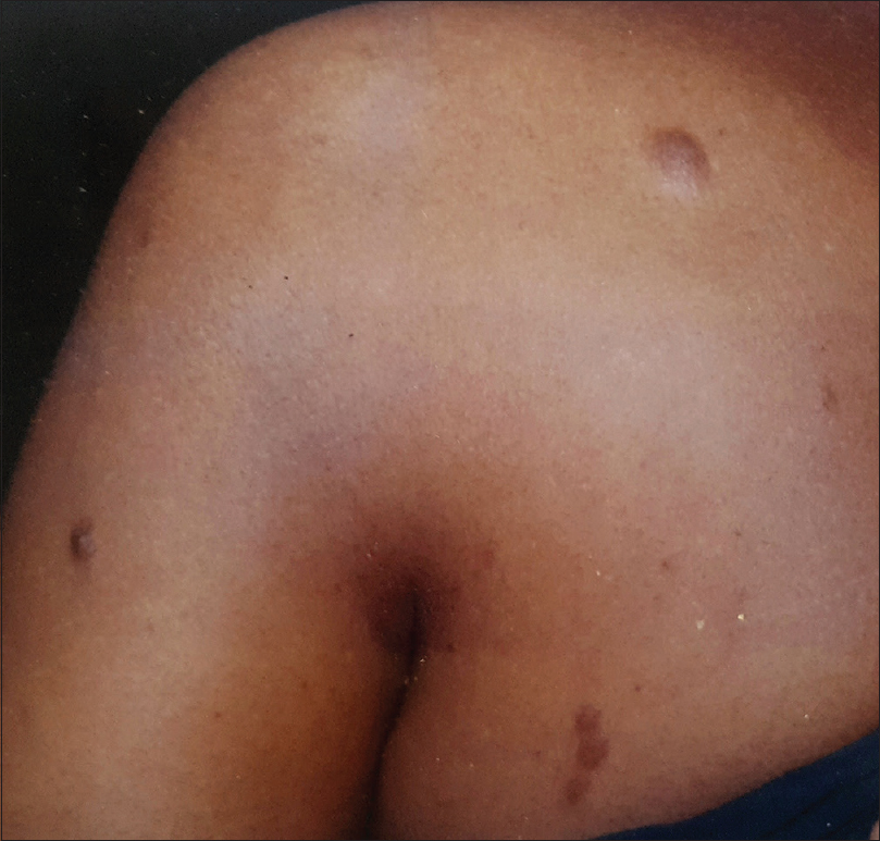 |
| Figure 1: Leiomyoma cutis: multiple, hyperpigmented, soft nodules over left shoulder, arm and upper back |
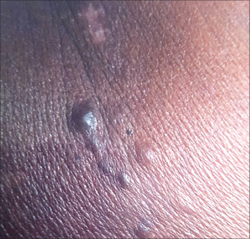 |
| Figure 2: Leiomyoma cutis: multiple skin-colored to hyperpigmented nodules with a few appearing translucent |
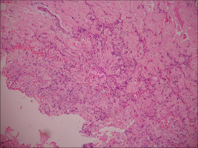 |
| Figure 3: Histopathology of leiomyoma: Dermis shows intersecting fascicles of smooth muscle bundles intermingled with collagen bundles in histopathology on high power (hematoxylin and eosin, ×400) |
Eccrine Spiradenoma
It presents as painful, blue-colored, small, nondescript lesions showing histologic differentiatiion towards intradermal eccrine ductal and secretory cells. Histopathology shows deeply basophilic stained, sharply marginated lobules lying freely in the dermis. Two types of epithelial cells are present – small cells with dark peripheral nuclei and pale cells with central nuclei.[8],[9],[10] Characteristic appearance is called “blue balls” in the dermis.[11]
Neuromas
These tumors present as painful skin colored lesions which are characterized by proliferative bundles of nerve fibres with a surrounding capsule on histopathology. Morton's neuroma presents as a reactive, persistent and degenerative nodule commonly over the sole.[4]
Dermatofibroma
It presents as a painful hyperpigmented nodule with “dimple sign” most commonly over the legs[4] [Figure - 4]. Histopathology shows a hyperplastic epidermis separated by a grenz zone from a dermal tumor composed of varying proportion of histiocytes and blood vessels [Figure - 5].[12]
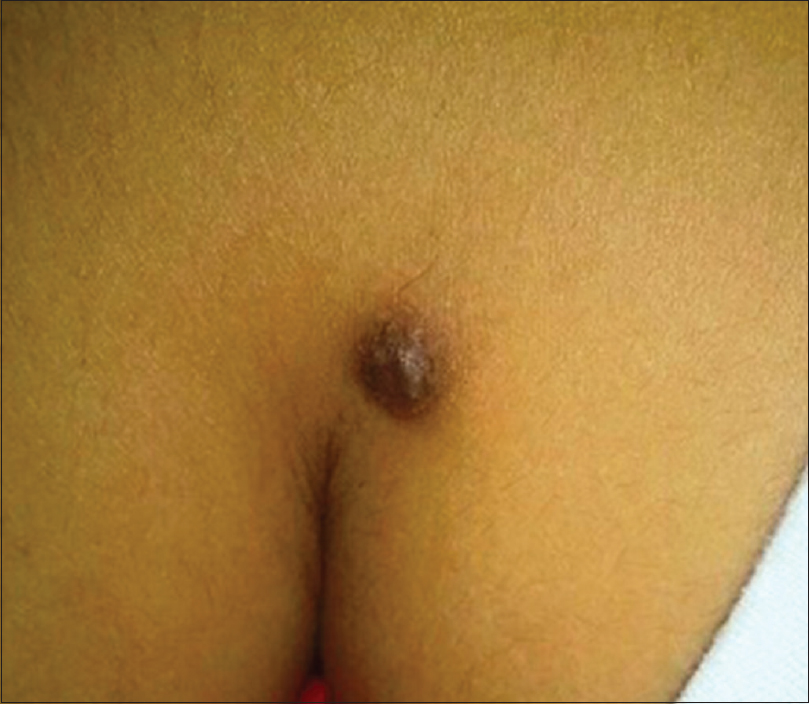 |
| Figure 4: Dermatofibroma: solitary, hyperpigmented firm nodule over upper back |
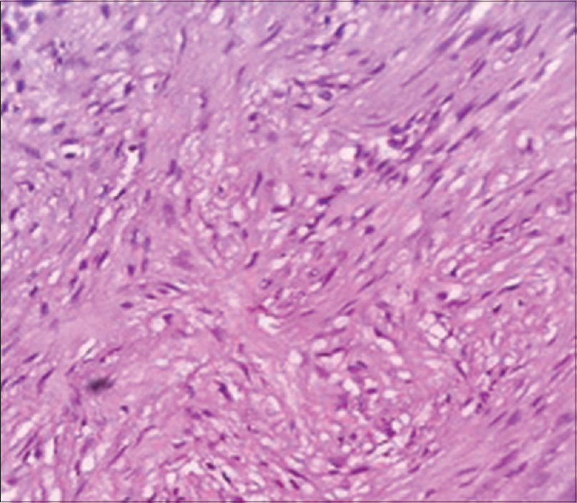 |
| Figure 5: Histopathology of dermatofibroma: Storiform pattern of spindle-shaped cells on high power (hematoxylin and eosin, ×400) |
Tufted Angiomas
Tufted angioma is an uncommon, benign, vascular tumor that usually develops during infancy or childhood, with a majority of lesions arising before the age of 5 years.[13] It typically presents on the neck, shoulders, trunk or groin as slowly expanding, mottled, red-to-purple patches and firm plaques superimposed with papules and nodules. Lesions are often associated with paroxysmal pain or tenderness to palpation and localized hyperhidrosis as well as hypertrichosis have been observed.[14] Kasabach–Merrit syndrome has been associated with congenital tufted angioma. Histopathologically, tufted angiomas are characterized by multiple, discrete lobules of tightly packed capillaries in a cannonball pattern within the dermis and sometimes in subcutis.[15]
Angiolipoma
It is a variant of lipoma.[4] Cutaneous angiomyolipomas are rare and differ from renal angiomyolipomas in that they are more common in males 33–77 years of age without any association with tuberous sclerosis and are HMB-45 negative. Histologically, they are composed of thick-walled blood vessels, smooth muscle cells and mature fat in variable proportions. Epithelioid cell component is usually absent in cutaneous angiomyolipomas in contrast to renal angiomyolipomas, which may be responsible for HMB-45 negativity.[16]
Neurilemmomas
These are nerve sheath tumors presenting as ovoid, rounded, firm, circumscribed nodules.[17] Neurilemmoma is one of the few truly encapsulated neoplasms of the human body.[18] Schwann cells are arranged in bands which stream and interweave. Cellular areas are known as Antoni A areas which are intermixed with areas showing predominantly myxoid change and named as Antoni B areas.[17]
Endometrioma
It is a rare skin tumor presenting with pain and bleeding which worsens at the time of menstruation. It usually appears over a scar or near umbilicus.[19] Histopathology shows a cellular vascular stroma with lumina as well as changes associated with various phases of menstrual cycle.[20]
Granular Cell Tumor
It occurs most commonly over the tongue. Histopathology shows irregular polygonal, large cells with poorly-defined membrane and granular cytoplasm.[17]
Glomus Tumor
This is a neoplasm of normal glomus body with a triad of symptoms of pain, pinpoint tenderness on blunt palpation and hypersensitivity to cold.[21] Commonly seen over digits but extradigital locations have also been described. We have reported a case of glomus tumor over the calf region [Figure - 6].[22] Histopathology shows branching vascular channels separated by connective tissue stroma containing aggregates, nests and masses of glomus cells [Figure - 7].[3]
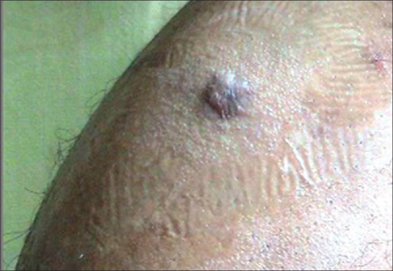 |
| Figure 6: Glomus tumor: dusky, cyanotic nodule over the left calf |
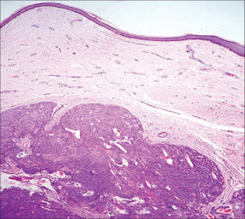 |
| Figure 7: Histopathology of glomus tumor: with hematoxylin and eosin stain showing a well-defined tumor in the dermis with solid sheets of tumor cells which are surrounding vascular channels (×100) |
Declaration of patient consent
The authors certify that they have obtained all appropriate patient consent forms. In the form, the patient has given his consent for his images and other clinical information to be reported in the journal. The patient understand that name and initials will not be published and due efforts will be made to conceal identity, but anonymity cannot be guaranteed.
Acknowledgment
The authors would like to acknowledge Dr. Sherina Laskar, MD, DNB, Department of Dermatology, Gauhati Medical College for her contribution towards reference articles for causes of painful tumors of the skin.
Financial support and sponsorship
Nil.
Conflicts of interest
There are no conflicts of interest.
[22-27]
| 1. |
Thappa DM, Garg BR, Prasad RR, Ratnakar C. Multiple leiomyoma cutis associated with Becker's nevus. J Dermatol 1996;23:719-20.
[Google Scholar]
|
| 2. |
Kamath GH, Bhat RM, Kumar S. Tufted angioma. Int J Dermatol 2005;44:1045-7.
[Google Scholar]
|
| 3. |
Bhat MR, George AA, Pinto AC, Sukumar D, Lyngdoh RH. Violaceous painful nodule of the leg in an Indian male patient. Indian J Dermatol Venereol Leprol 2012;78:410.
[Google Scholar]
|
| 4. |
Naversen DN, Trask DM, Watson FH, Burket JM. Painful tumors of the skin: “LEND AN EGG”. J Am Acad Dermatol 1993;28:298-300.
[Google Scholar]
|
| 5. |
James WD, Berger TG, Elston DM. Dermal and subcutaneous tumors. In: Andrews' Diseases of Skin: Clinical Dermatology. 10th ed. Philadelphia: W. B. Saunders; 2006. p. 584-5.
[Google Scholar]
|
| 6. |
Fisher WC, Helwig EB. Leiomyomas of the skin. Arch Dermatol 1963;88:510-20.
[Google Scholar]
|
| 7. |
Enzinger FM, Weiss SW, editors. Soft Tissue Tumors. St. Louis: The C. V. Mosby Co.; 1983. p. 583-5.
[Google Scholar]
|
| 8. |
Kaleeswaran AV, Janaki VR, Sentamilselvi G, Kiruba MC. Eccrine spiradenoma. Indian J Dermatol Venereol Leprol 2002;68:236-7.
[Google Scholar]
|
| 9. |
Kersting DW, Helwig EB. Eccrine spiradenoma. AMA Arch Derm 1956;73:199-227.
[Google Scholar]
|
| 10. |
Mambo NC. Eccrine spiradenoma: Clinical and pathologic study of 49 tumors. J Cutan Pathol 1983;10:312-20.
[Google Scholar]
|
| 11. |
Klein W, Chan E, Seykora JT. Tumors of the epidermal appendages. In: Elder DE, Rosalie E, Johnson BL, Murphy GF, editors. Levers Histopathology of the Skin. 10th ed. India: Lippincott Williams & Wilkins; 2009. p. 904.
[Google Scholar]
|
| 12. |
Heenan PJ. Tumors of fibrous tissue involving the skin. In: Elder DE, Rosalie E, Johnson BL, Murphy GF, editors. Levers Histopathology of the Skin. 10th ed. India: Lippincott Williams & Wilkins; 2009. p. 979.
[Google Scholar]
|
| 13. |
Jones EW, Orkin M. Tufted angioma (angioblastoma). A benign progressive angioma, not to be confused with Kaposi's sarcoma or low-grade angiosarcoma. J Am Acad Dermatol 1989;20:214-25.
[Google Scholar]
|
| 14. |
Herron MD, Coffin CM, Vanderhooft SL. Tufted angiomas: Variability of the clinical morphology. Pediatr Dermatol 2002;19:394-401.
[Google Scholar]
|
| 15. |
Arai E, Kuramochi A, Tsuchida T, Tsuneyoshi M, Kage M, Fukunaga M, et al. Usefulness of D2-40 immunohistochemistry for differentiation between kaposiform hemangioendothelioma and tufted angioma. J Cutan Pathol 2006;33:492-7.
[Google Scholar]
|
| 16. |
Val-Bernal JF, Mira C. Cutaneous angiomyolipoma. J Cutan Pathol 1996;23:364-8.
[Google Scholar]
|
| 17. |
Calonje E. Soft tissue tumors and tumor like conditions. In: Burns T, Bethnach S, Cox N, Griffiths C, editors. Rook's Textbook of Dermatology. 8th ed. Oxford: Blackwell Science; 2010. p. 56.50
[Google Scholar]
|
| 18. |
Rodrigues G, Rao A. Digital neurilemmoma: A case report. Bahrein Med Bull 2002;24:1-3.
[Google Scholar]
|
| 19. |
Steck WD, Helwig EB. Cutaneous endometriosis. JAMA 1965;191:167-70.
[Google Scholar]
|
| 20. |
Tidman MJ, Macdonald DM. Cutaneous endometriosis: A histopathological study. J Am Acad Dermatol 1988;18:373-7.
[Google Scholar]
|
| 21. |
Schiefer TK, Parker WL, Anakwenze OA, Amadio PC, Inwards CY, Spinner RJ, et al. Extradigital glomus tumors: A 20-year experience. Mayo Clin Proc 2006;81:1337-44.
[Google Scholar]
|
| 22. |
Perks FJ, Beggs I, Lawson GM, Davie R. Juxtacortical glomus tumor of the distal femur adjacent to the popliteal fossa. AJR Am J Roentgenol 2003;181:1590-2.
[Google Scholar]
|
| 23. |
Jin XL, Wang ZH, Xiao XB, Huang LS, Zhao XY. Blue rubber bleb nevus syndrome: A case report and literature review. World J Gastroenterol 2014;20:17254-9.
[Google Scholar]
|
| 24. |
Park HR, Im SB, Kim HK, Shin DS, Park YL. Painful eccrine spiradenoma containing nerve fibers: A case report. Dermatology 2012;224:301-6.
[Google Scholar]
|
| 25. |
Lee WJ, Jung JM, Won CH, Chang SE, Choi JH, Moon KC, et al. Clinical and histological patterns of dermatofibroma without gross skin surface change: A comparative study with conventional dermatofibroma. Indian J Dermatol Venereol Leprol 2015;81:263-9.
[Google Scholar]
|
| 26. |
Bernstein EF, Kantor G, Howe N, Savit RM, Koblenzer PJ, Uitto J, et al. Tufted angioma of the thigh. J Am Acad Dermatol 1994;31:307-11.
[Google Scholar]
|
| 27. |
Heimburg DV, Biesterfeld S, Aslani A, Pallua N. Painful angiolipomatosis: Evaluation of the extended subcutaneous lipectomy and immunohistochemical and histological analysis compared to other benign lipomatous tumors. Eur J Plast Surg 2003;25:396-400.
[Google Scholar]
|
Fulltext Views
25,687
PDF downloads
7,030


