Translate this page into:
Pemphigus vegetans localized to unusual sites
2 Department of Histopathology, Postgraduate Institute of Medical Education and Research, Chandigarh, India
Correspondence Address:
Dipankar De
Department of Dermatology, Venereology, and Leprology, Postgraduate Institute of Medical Education and Research, Chandigarh - 160 012
India
| How to cite this article: Khullar G, De D, Narang T, Saikia UN, Handa S. Pemphigus vegetans localized to unusual sites. Indian J Dermatol Venereol Leprol 2015;81:509-511 |
Sir,
Pemphigus vegetans is an uncommon variant of pemphigus vulgaris that commonly manifests as verrucous vegetating plaques in the flexures. We describe a case of pemphigus vegetans (Hallopeau type) localized to atypical sites with no mucosal involvement. A high index of suspicion is required to diagnose pemphigus vegetans on non-intertriginous sites and diagnosis is confirmed by histopathological and immunological studies.
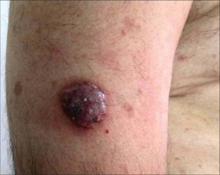
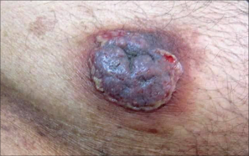
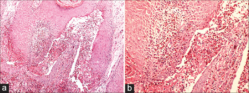
A 64-year-old man presented with itchy, erythematous verrucous plaques on the right arm, thigh and abdomen for 3 months. The lesions started as pustules, which ruptured to form erosions and gradually enlarged to develop into elevated plaques. On physical examination, well-defined, hypertrophic, vegetating, dusky red plaques measuring 1.5 × 1.5, 2 × 1.5 and 3 × 2.5 cm, with peripheral vesico-pustules and erosions were present on the right arm, posterior aspect of right thigh and lower abdomen, respectively. The lesions were surrounded by a rim of erythema [Figure - 1] and [Figure - 2]. Mucous membranes were spared. A provisional clinical diagnosis of pemphigus vegetans was considered. Laboratory investigations revealed peripheral blood eosinophilia (eosinophils 10%, normal range: 1-5% of the differential leukocyte count). Tzanck smear from the lesion showed acantholytic cells. Histpathological examination from the abdominal lesion showed hyperkeratosis, acanthosis, papillomatosis and a suprabasal blister containing an eosinophil-rich infiltrate forming microabscesses in the epidermis. Upper dermis showed a perivascular infiltrate with predominant eosinophils [Figure - 3]a and b. Direct immunofluorescence studies showed intercellular IgG deposits (3+) in the epidermis. Hence the diagnosis of pemphigus vegetans (Hallopeau type) was confirmed. The patient was started on oral prednisolone, 20 mg once a day and azathioprine 50 mg twice a day. He showed about 75% improvement after 1 month, following which oral prednisolone was gradually tapered and stopped over the next 2 months and azathioprine was continued as maintenance therapy. The patient has been followed up for 5 months and has developed no new lesions, while the older lesions have healed with the formation of acanthomas.
 |
| Figure 1: Well-defined, erythematous, verrucous plaque with vesico-pustules on the periphery on the right arm |
 |
| Figure 2: Dusky red, hypertrophic plaque showing peripheral rim of vesico-pustules with erosions at places on the lower abdomen |
 |
| Figure 3: (a) The epidermis shows acanthosis and a suprabasal cleft containing numerous eosinophils forming microabscesses. The superficial dermis shows a perivascular infiltrate rich in eosinophils (H and E, ×100), (b) Higher magnification depicting eosinophilic microabscesses within the suprabasal split and the dermal papillae (H and E, ×200) |
Pemphigus vegetans is an uncommon variant of pemphigus vulgaris that contributes to 5% of all pemphigus cases. [1] In a recent retrospective study (1981-2009) of pemphigus vegetans from Tunisia, the prevalence rate was 9.1% among all patients of pemphigus. [2] It is categorized into two subtypes based on the initial presentation and disease course: Hallopeau and Neumann. The former begins as pustules and has a relatively benign course while the later, which is more frequent starts as flaccid vesicles and bullae and shows a poor response to therapy. Both the forms gradually evolve into hypertrophic vegetating plaques predominantly distributed in the intertriginous sites, although any site may be involved. Oral mucosa is affected in nearly 60-80% cases and often at the onset of the disease. Less commonly, pemphigus vegetans may be localized to unusual sites such as scalp, face, breast, leg and sole. [2],[3],[4] Diagnosis poses a challenge when the disease is limited to non-intertriginous sites and requires a high index of suspicion. Differential diagnoses include pyodermatitis-pyostomatitis vegetans, vegetative pyoderma gangrenosum, pemphigoid vegetans, blastomycosis-like pyoderma and halogenoderma. Pyodermatitis-pyostomatitis vegetans simulates pemphigus vegetans, both clinically and histologically, but classically shows negative immunofluorescence. However, intercellular IgA deposition in pyodermatitis-pyostomatitis vegetans has been reported and may be regarded as an epiphenomenon secondary to epithelial damage or representing a pemphigus vegetans variant of IgA pemphigus. [5] Most patients with pyodermatitis-pyostomatitis vegetans have an underlying inflammatory bowel disease. Vegetative pyoderma gangrenosum is a rare, superficial, non-aggressive variant of pyoderma gangrenosum that usually lacks association with any systemic disease. It commonly presents on the trunk as an abscess or a nodule that gradually develops into a vegetative plaque with sinus tracts. Histology reveals sinus formation, neutrophilic abscesses and palisading three layered granulomas. Pemphigoid vegetans is an extremely uncommon variant of bullous pemphigoid that can be differentiated from pemphigus vegetans on histology and immunofluorescence. The principal autoantigen in pemphigus vegetans is desmoglein (Dsg) 3, although in some cases Dsg1 and desmocollin 1, 2 and 3 have also been implicated. Histopathologically, early pustular lesions of Hallopeau type demonstrate eosinophilic spongiosis with transmigration of eosinophils into the epidermis and eosinophilic microabscesses as seen in our case, while Neumann type shows suprabasal vesicles with acantholytic cells but no eosinophilic microabscesses. Chronic vegetative lesions in both subtypes reveal hyperkeratosis, acanthosis, papillomatosis and a suprabasal cleft with eosinophils. Direct immunofluorescence shows IgG deposits usually associated with C3 on the keratinocyte cell surface; however, coexistence of IgA anti-Dsg3 antibodies has also been described. [6] Systemic corticosteroids are considered the mainstay of treatment of pemphigus vegetans. Adjuvant immunosuppressants and immunomodulatory agents including azathioprine, cyclophosphamide, cyclosporine, intravenous immunoglobulin, nicotinamide with tetracycline, dapsone, and retinoids may be required to enhance the treatment response. [2] Our patient responded well to oral prednisolone and azathioprine, consistent with the expected clinical behavior of Hallopeau type. The present case was unusual as the lesions were non-flexural in distribution and oral mucosa was not involved. The eosinophilic microabscesses seen on histopathology corresponded to the pustules in the periphery of the hypertrophic plaques, thereby confirming the diagnosis of pemphigus vegetans (Hallopeau type).
| 1. |
Becker BA, Gaspari AA. Pemphigus vulgaris and vegetans. Dermatol Clin 1993;11:429-52.
[Google Scholar]
|
| 2. |
Zaraa I, Sellami A, Bouguerra C, Sellami MK, Chelly I, Zitouna M, et al. Pemphigus vegetans: A clinical, histological, immunopathological and prognostic study. J Eur Acad Dermatol Venereol 2011;25:1160-7.
[Google Scholar]
|
| 3. |
Wei J, He CD, Wei HC, Li B, Wang YK, Jin GY, et al. Facial pemphigus vegetans. J Dermatol 2011;38:615-8.
[Google Scholar]
|
| 4. |
Jain V, Jindal N, Imchen S. Localized pemphigus vegetans without mucosal involvement. Indian J Dermatol 2014;59:210.
[Google Scholar]
|
| 5. |
Wolz MM, Camilleri MJ, McEvoy MT, Bruce AJ. Pemphigus vegetans variant of IgA pemphigus, a variant of IgA pemphigus and other autoimmune blistering disorders. Am J Dermatopathol 2013;35:e53-6.
[Google Scholar]
|
| 6. |
Morizane S, Yamamoto T, Hisamatsu Y, Tsuji K, Oono T, Hashimoto T, et al. Pemphigus vegetans with IgG and IgA antidesmoglein 3 antibodies. Br J Dermatol 2005;153:1236-7.
[Google Scholar]
|
Fulltext Views
4,953
PDF downloads
2,141


