Translate this page into:
Peutz-Jeghers syndrome with prominent palmoplantar pigmentation
Correspondence Address:
K N Shivaswamy
Department of Dermatology and STD, M. S. Ramaiah Medical College, Bangalore - 560 054, Karnataka
India
| How to cite this article: Shivaswamy K N, Shyamprasad A L, Sumathy T K, Ranganathan C. Peutz-Jeghers syndrome with prominent palmoplantar pigmentation. Indian J Dermatol Venereol Leprol 2008;74:154-155 |
 |
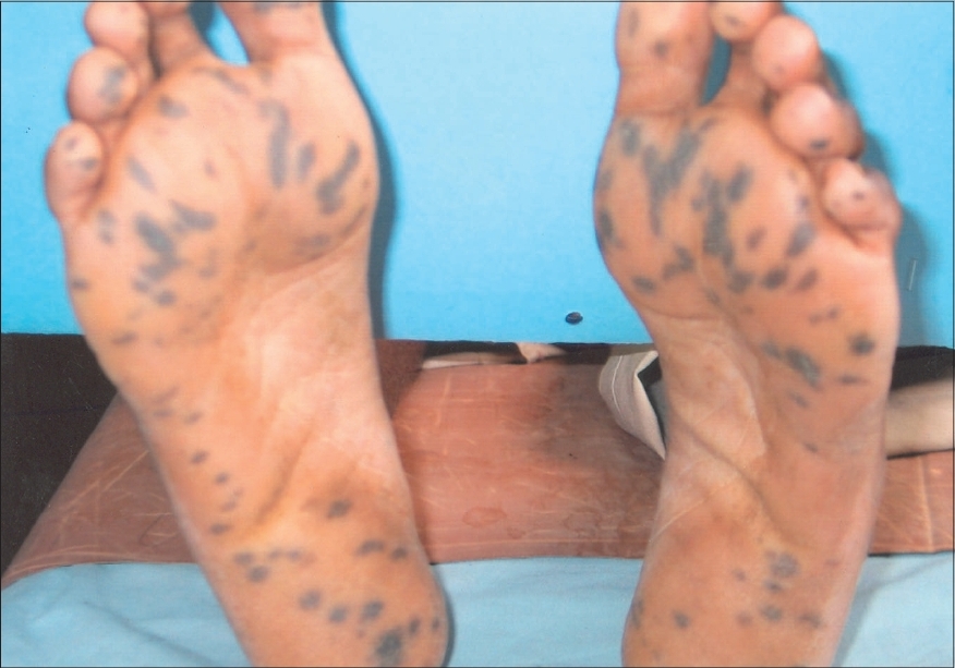
| Figure 2: Large hyperpigmented macules over the soles |
 |
| Figure 2: Large hyperpigmented macules over the soles |
 |
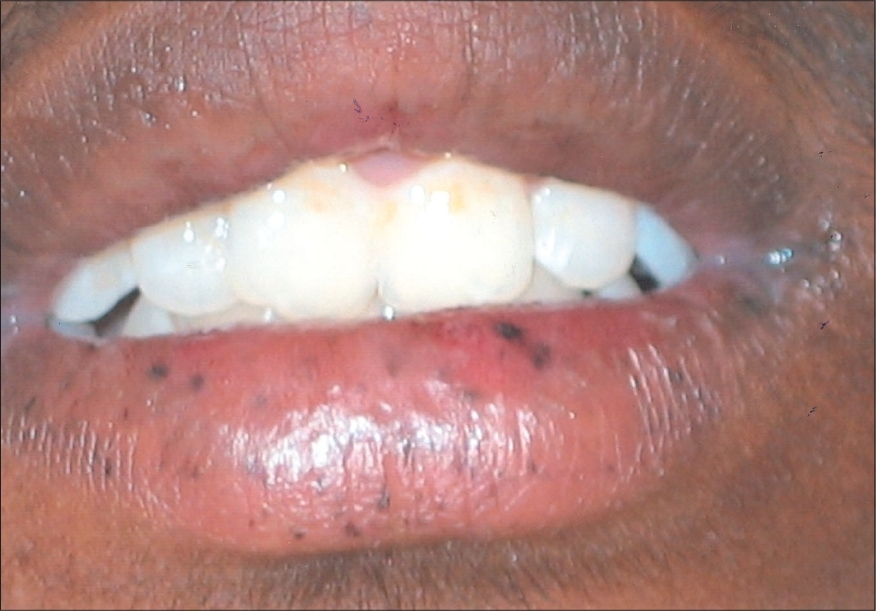
| Figure 1: Photograph showing lentigenes over the lips |
 |
| Figure 1: Photograph showing lentigenes over the lips |
Sir,
Peutz-Jeghers syndrome (PJS) is an autosomal dominant disorder characterized by lentigines on the lips, buccal mucosa, tips of fingers and toes in association with intestinal polyps. PJS is caused by mutations in the gene coding for serine threonine kinase, located on the p arm of chromosome 19. [1],[2] The pigmented macules that usually appear during infancy and early childhood, are seen over the back of the hands and tips of the fingers and toes but are very rarely seen over the palms and soles. [3]
A 17-year-old male was referred from the surgery department to us for evaluation of the pigmented lesions over his palms and soles. Recently, he was operated for persistent colicky abdominal pain and was found to have an exophytic growth in the jejunum, which showed evidence of villous adenomas on histopathological examination. On enquiry, the patient gave a history of pigmented lesions over the palms and soles as well as the lips since childhood that increased in size and number over the past year. Two of his siblings also had history of pigmentation of the lips as well as their palms and soles. On examination, multiple discrete-to-coalescent, hyperpigmented macules 0.5-1.5 mm in size were seen over the lips [Figure - 1], perioral area and buccal mucosa. Similar hyperpigmented and round-to-oval macules of varying sizes ranging from 3 mm to more than 1 cm in diameter were present over the tips of the fingers and up to 5 cm in diameter were seen over the soles [Figure - 2]. A 12 cm-long paramedian linear scar was evident over the abdomen. A diagnosis of Peutz-Jeghers syndrome was made.
The pigmented macules of Peutz-Jeghers syndrome (PJS) usually appear during infancy and early childhood, but may be present at birth or develop later in life and have a tendency to increase in size during adolescence. Round, oval or irregular patches of brown or almost black pigmentation, 1-5 mm in diameter, are irregularly distributed over the buccal mucosa, gums, hard palate and lips, especially the lower lip. The pigmented macules may also occur on the face especially around the nose and the mouth but are smaller and often under 1 mm in size. Large macules are rarely seen over the back of the hands and the tips of the fingers and toes. [3] Our case had large hyperpigmented macules over both his palms and soles [Figure - 2], which is very unusual.
Intestinal manifestations include numerous polyps in the jejunum, ileum and less frequently in the colon, rectum, stomach and duodenum. Patients with PJS have a 10-18 fold greater lifetime cancer risk than the general population. The greatest risk is for gastrointestinal malignancy of the colon and duodenum. [4] Laugier-Hunziker syndrome and Cronkhite-Canada syndrome should be considered in the differential diagnosis of PJS. In the former, mucosal and nail pigmentation are characteristic but without intestinal polyps, whereas the volar aspects of the fingers are heavily pigmented along with evidence of intestinal polyps but without mucosal pigmentation in the latter. [5],[6]
Although perioral pigmentation is commonly seen in PJS, palmoplantar pigmentation is rare and such numerous large hyperpigmented macules over the soles have never been described before and hence, being reported.[7]
| 1. |
Dormandy TL. Gastrointestinal polyposis with mucocutaneous pigmentation (Peutz-Jeghers syndrome). N Engl J Med 1949;241:993-1005.
[Google Scholar]
|
| 2. |
Tsao H. Update on familial cancer syndromes and the skin. J Am Acad Dermatol 2000;42:939-46.
[Google Scholar]
|
| 3. |
Bleehen SS, Anstery AV. Disorders of skin colour. In : Rook's text book of dermatology. Burns T, Breathnach S, Cox N, Griffiths C, editors. 7 th ed. Oxford: Blackwell Science; 2007. p. 1-39.68.
th ed. Oxford: Blackwell Science; 2007. p. 1-39.68.'>[Google Scholar]
|
| 4. |
Disturbances of pigmentation. In : Andrew's diseases of the skin. Clinical Dermatology. James WD, Berger TG, Elston DM, editors. 10 th ed. Philadelphia: Saunders-Elsevier; 2006. p. 853-68.
th ed. Philadelphia: Saunders-Elsevier; 2006. p. 853-68.'>[Google Scholar]
|
| 5. |
Veraldi S, Cavicchini S, Benelli C, Gasparini G. Laugier-Hunziker syndrome: A clinical, histopathologic and ultrastructural study of four cases and review of the literature. J Am Acad Dermatol 1991;25:632-6.
[Google Scholar]
|
| 6. |
Herzberb AJ, Kaplan DL. Cronkhite-Canada syndrome. Int J Dermatol 1990;29:121-5.
[Google Scholar]
|
| 7. |
Ortonne J, Bahadoran P, Fitzpatric TB, Mosher DB, Hori Y. Hypomelanosis and hypermelanosis. In : Fitzpatric's dermatology in general medicine. Freedberg IM, Eisener AZ, Wolff, et al , editors. 6 th ed, McGraw-Hill: 2003. p. 836-81.
et al , editors. 6 th ed, McGraw-Hill: 2003. p. 836-81.'>[Google Scholar]
|
Fulltext Views
4,665
PDF downloads
1,864


