Generic selectors
Exact matches only
Search in title
Search in content
Post Type Selectors
Search in posts
Search in pages
Filter by Categories
15th National Conference of the IAOMFP, Chennai, 2006
Abstract
Abstracts from current literature
Acne in India: Guidelines for management - IAA Consensus Document
Addendum
Announcement
Art & Psychiatry
Article
Articles
Association Activities
Association Notes
Award Article
Book Review
Brief Report
Case Analysis
Case Letter
Case Letters
Case Notes
Case Report
Case Reports
Clinical and Laboratory Investigations
Clinical Article
Clinical Studies
Clinical Study
Commentary
Conference Oration
Conference Summary
Continuing Medical Education
Correspondence
Corrigendum
Cosmetic Dermatology
Cosmetology
Current Best Evidence
Current Issue
Current View
Derma Quest
Dermato Surgery
Dermatopathology
Dermatosurgery Specials
Dispensing Pearl
Do you know?
Drug Dialogues
e-IJDVL
Editor Speaks
Editorial
Editorial Remarks
Editorial Report
Editorial Report - 2007
Editorial report for 2004-2005
Errata
Erratum
Focus
Fourth All India Conference Programme
From Our Book Shelf
From the Desk of Chief Editor
General
Get Set for Net
Get set for the net
Guest Article
Guest Editorial
History
How I Manage?
IADVL Announcement
IADVL Announcements
IJDVL Awards
IJDVL AWARDS 2015
IJDVL Awards 2018
IJDVL Awards 2019
IJDVL Awards 2020
IJDVL International Awards 2018
Images in Clinical Practice
Images in Dermatology
In Memorium
Inaugural Address
Index
Knowledge From World Contemporaries
Leprosy Section
Letter in Response to Previous Publication
Letter to Editor
Letter to the Editor
Letter to the Editor - Case Letter
Letter to the Editor - Letter in Response to Published Article
LETTER TO THE EDITOR - LETTERS IN RESPONSE TO PUBLISHED ARTICLES
Letter to the Editor - Observation Letter
Letter to the Editor - Study Letter
Letter to the Editor - Therapy Letter
Letter to the Editor: Articles in Response to Previously Published Articles
Letters in Response to Previous Publication
Letters to the Editor
Letters to the Editor - Letter in Response to Previously Published Articles
Letters to the Editor: Case Letters
Letters to the Editor: Letters in Response to Previously Published Articles
Media and news
Medicolegal Window
Messages
Miscellaneous Letter
Musings
Net Case
Net case report
Net Image
Net Images
Net Letter
Net Quiz
Net Study
New Preparations
News
News & Views
Obituary
Observation Letter
Observation Letters
Oration
Original Article
ORIGINAL CONTRIBUTION
Original Contributions
Pattern of Skin Diseases
Pearls
Pediatric Dermatology
Pediatric Rounds
Perspective
Presedential Address
Presidential Address
Presidents Remarks
Quiz
Recommendations
Regret
Report
Report of chief editor
Report of Hon : Treasurer IADVL
Report of Hon. General Secretary IADVL
Research Methdology
Research Methodology
Resident page
Resident's Page
Resident’s Page
Residents' Corner
Residents' Corner
Residents' Page
Retraction
Review
Review Article
Review Articles
Reviewers 2022
Revision Corner
Self Assessment Programme
SEMINAR
Seminar: Chronic Arsenicosis in India
Seminar: HIV Infection
Short Communication
Short Communications
Short Report
Snippets
Special Article
Specialty Interface
Studies
Study Letter
Study Letters
Supplement-Photoprotection
Supplement-Psoriasis
Symposium - Contact Dermatitis
Symposium - Lasers
Symposium - Pediatric Dermatoses
Symposium - Psoriasis
Symposium - Vesicobullous Disorders
SYMPOSIUM - VITILIGO
Symposium Aesthetic Surgery
Symposium Dermatopathology
Symposium-Hair Disorders
Symposium-Nails Part I
Symposium-Nails-Part II
Systematic Review and Meta-Analysis
Systematic Reviews and Meta-analyses
Systematic Reviews and Meta-analysis
Tables
Technology
Therapeutic Guideline-IADVL
Therapeutic Guidelines
Therapeutic Guidelines - IADVL
Therapeutics
Therapy
Therapy Letter
Therapy Letters
View Point
Viewpoint
What’s new in Dermatology
View/Download PDF

Translate this page into:

Observation Letter
ARTICLE IN PRESS
doi:
10.25259/IJDVL_1325_2023

Reticulate sclerodermoid pigmentation in Hutchinson–Gilford progeria syndrome – A report of two cases

Department of Dermatology and Venereology, All India Institute of Medical Sciences, Bhubaneswar, Odisha, India
Department of Dermatology, Venereology and Leprology, Post Graduate Institute of Medical Education and Research, Chandigarh, India
Department of Pathology and Lab Medicine, All India Institute of Medical Sciences, Bhubaneswar, Odisha, India.

Corresponding author: Dr. Vishal Thakur, Department of Dermatology and Venereology, All India Institute of Medical Sciences, Bhubaneswar, Odisha, India. drvishal87igmc@gmail.com

Licence
This is an open-access article distributed under the terms of the Creative Commons Attribution-Non Commercial-Share Alike 4.0 License, which allows others to remix, transform, and build upon the work non-commercially, as long as the author is credited and the new creations are licensed under the identical terms.

How to cite this article: Gowda SK, Baskaran N, Behera B, Ayyanar P, Vinay K, Thakur V. Reticulate sclerodermoid pigmentation in Hutchinson-Gilford progeria syndrome – A report of two cases. Indian J Dermatol Venereol Leprol. doi: 10.25259/IJDVL_1325_2023

Dear Editor,

Hutchinson–Gilford Progeria syndrome (HGPS, OMIM 176670) is a rare (prevalence 1 in 4 million births) autosomal dominant genetic premature ageing disorder due to mutation in the lamin A (LMNA) gene.1 Only about 150 cases of this disease have been reported to date. It has characteristic facial features such as a disproportionately larger head than face, delayed dentition, loss of scalp hair, delayed sexual maturity and cardiovascular abnormalities.2 Herein, we present two cases of HGPS having the unique feature of reticulate hyperpigmentation on the trunk with sclerodermoid skin changes and fat outpouching.

Our index case was a 14-month-old boy born of a non-consanguineous marriage who presented with loss of hair on the scalp and pigmentation on the abdomen, back and lower limbs since 1 month of age. There was no history of similar features among siblings or first-degree relatives. On examination, the height (73cm) and weight (7.5 kg) of the child were between the 3rd and 50th centile for his age. The face showed frontal bossing, prominent scalp veins, a sculptured nose, midfacial hypoplasia, micrognathia, protruded eyes and ears with a thin helix and wide concha [Figure 1a]. Interestingly, the child had in addition, reticulate pigmentation on the abdomen, back and lower limbs; along with binding down of abdominal skin with pebble-like outpouchings from the abdominal wall [Figure 1b]. Biopsy from the area having reticulate hyperpigmentation and outpouching of skin revealed epidermal acanthosis, increased basal layer melanin and melanophages in the superficial dermis with diffuse dermal sclerosis [Figure 2]. Whole exome sequencing confirmed the clinical suspicion of HGPS with heterozygous variation in Exon 11 of the LMNA gene in the form of single base substitution at 1824 nucleotide sequence (c.1824C>T).
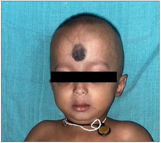
Case 1 showing characteristic facial features including frontal bossing, sparse eyebrows and eyelashes, flat and broad nasal bridge, prominent philtrum, micrognathia and circumoral cyanosis, sparse and thin hair and prominent dilated veins visible through the translucent skin.
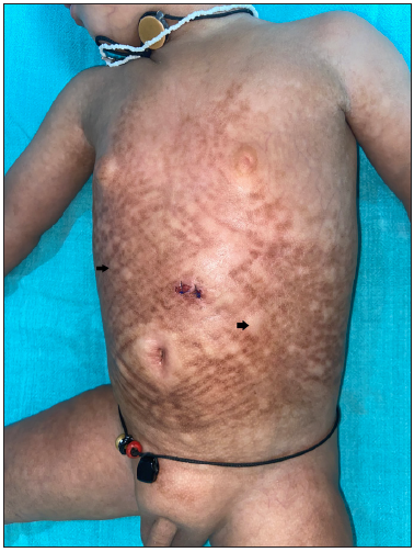
Figure 1a:
Case 1 showing characteristic facial features including frontal bossing, sparse eyebrows and eyelashes, flat and broad nasal bridge, prominent philtrum, micrognathia and circumoral cyanosis, sparse and thin hair and prominent dilated veins visible through the translucent skin.
Sclerodermoid changes in reticulate pattern along with mottled hyperpigmentation on the abdomen. Pebble-like outpouchings of fat on the abdominal wall (black arrows).
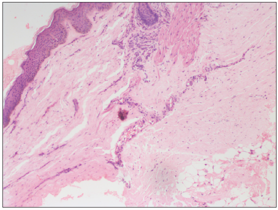
Figure 1b:
Sclerodermoid changes in reticulate pattern along with mottled hyperpigmentation on the abdomen. Pebble-like outpouchings of fat on the abdominal wall (black arrows).
Epidermal acanthosis, increased basal layer pigmentation and melanophages in the superficial dermis with diffuse dermal sclerosis (Haematoxylin & Eosin, 100x).
Figure 2:
Epidermal acanthosis, increased basal layer pigmentation and melanophages in the superficial dermis with diffuse dermal sclerosis (Haematoxylin & Eosin, 100x).

Another nine-month-old girl child presented with similar cutaneous findings to our index case [Figure 3]. Whole-exome sequencing revealed a pathogenic mutation in the LMNA gene at Exon 11 variant c. 1822G> A. The rest of the systemic examinations, including ocular, hearing and cardiac examination were normal in both children.
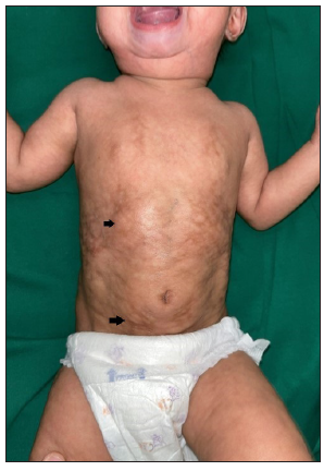
Case 2 showing sclerodermoid skin changes, mottled hyperpigmentation and fat outpouchings (black arrows) on the abdomen.
Figure 3:
Case 2 showing sclerodermoid skin changes, mottled hyperpigmentation and fat outpouchings (black arrows) on the abdomen.

HGPS is caused by abnormal lamin-A precursor progerin with retained farnesyl group leading to genomic instability and premature cell senescence.2 It is characterised by alopecia (100%), sclerodermoid skin changes (78%), prominent skin vasculature visible through translucent skin (22%), dyspigmentation (16%) and characteristic facies (excessive folding on forehead and cheeks, small ear lobes, protrusion of eyes, thin nasal bridge and collapsed flattened nasal tip).3 Our cases had reticulate pigmentation along with sclerodermoid skin changes prominent on the abdomen and proximal extremities.

Sclerodermoid changes in progeria initially (6 to 12 months) start as swollen and thick skin over the abdomen and proximal limbs which later (18 months) become sclerodermatous and bound down. As the disease progresses (around 2 years), the binding down vanishes and the skin becomes thin, atrophic and dry, with/without scaling.1 In a case series, the bilateral nipples and labium majus were spared during the initial sclerotic process, leading to a ‘protuberant soft nipples and labium majus’ which was labelled as ‘protuberant bikini sign’ (88.9%).4 However, in our cases, we noticed characteristic reticulate sclerodermoid hyperpigmented skin changes with outpouching of fat in uninvolved sclerotic areas giving a ‘plastic bubble wrap’ appearance.

Histopathology of sclerodermatous skin during the acute phase reveals normal epidermis, hypertrophic collagen and decreased fibroblast with scattered lymphocytes that invade the subcutaneous tissue. At a later stage, biopsy reveals thickened dermis and homogenised collagen in the deeper dermis with decreased cellularity. At the end stage, the biopsy shows replacement of the corium by fibrotic hyaline tissue, decreased sweat glands and sebaceous glands and atrophic subcutaneous adipose tissue.2

It is a fatal disease with most patients not surviving beyond 13 years due to death from stroke or myocardial infarction because of severe atherosclerosis. Management is based on counselling the parents regarding the disease prognosis and the need for regular follow-up to detect cardiovascular and skeletal abnormalities. Low-dose aspirin has been advocated as prophylaxis to delay atherosclerotic changes. FDA has recently approved lonafarnib (farnesyl transferase inhibitor) for this disorder offering hope in the management of this deadly disease.5 The drug however is not yet commercially available across many countries.

We report these cases to highlight the under-reported but characteristic cutaneous finding of reticulate sclerodermoid pigmentation and fat outpouchings in patients of HGPS. This morphological finding can help as a cutaneous marker of HGPS in resource-limited settings where facilities for genetic diagnosis are not easily available.

Declaration of patient consent

The authors certify that they have obtained all appropriate patient consent.

Financial support and sponsorship

Nil.

Conflicts of interest

There are no conflicts of interest.

Use of artificial intelligence (AI)-assisted technology for manuscript preparation

The authors confirm that there was no use of artificial intelligence (AI)-assisted technology for assisting in the writing or editing of the manuscript and no images were manipulated using AI.

References

  1. , , , . Hutchinson-Gilford Progeria Syndrome: A premature aging disease. Mol Neurobiol. 2018;55:4417-27.
    [CrossRef] [PubMed] [Google Scholar]
  2. . Hutchinson-Gilford progeria syndrome: Review of the phenotype. Am J Med Genet A. 2006;140:2603-24.
    [CrossRef] [PubMed] [Google Scholar]
  3. , , , , , . Initial cutaneous manifestations of Hutchinson-Gilford progeria syndrome. Pediatr Dermatol. 2014;31:196-202.
    [CrossRef] [PubMed] [PubMed Central] [Google Scholar]
  4. , , , , , . Clinical and genetic features of children with Hutchinson-Gilford progeria syndrome: A case series and a literature review. J Eur Acad Dermatol Venereol. 2021;35:e387-e91.
    [CrossRef] [PubMed] [Google Scholar]
  5. . Lonafarnib: First approval. Drugs. 2021;81:283-9.
    [CrossRef] [PubMed] [PubMed Central] [Google Scholar]

Fulltext Views
1,510

PDF downloads
2,268
View/Download PDF
Download Citations
BibTeX
RIS
Show Sections