Translate this page into:
Pigmented oral lesion in a patient with metastatic melanoma
2 Department of Pathology, Clinical Management Unit, University Hospital Puerta del Mar, Andalusian Health Service, Cádiz, Spain
Correspondence Address:
Cintia Arjona-Aguilera
Servicio Andaluz De Salud, Avenida Ana de Viya 21, 11009 Cádiz
Spain
| How to cite this article: Arjona-Aguilera C, Collantes-Rodríguez C, Gil-Jassogne C, Ossorio-García L, Jiménez-Gallo D. Pigmented oral lesion in a patient with metastatic melanoma. Indian J Dermatol Venereol Leprol 2018;84:117-119 |
Clinical History
A 64-year-old Caucasian man with metastatic melanoma of unknown origin was referred to the dermatology unit in a bid to detect the primary tumour. He had no family history of skin cancer or multiple nevi. Two right axillary lymph node metastatic masses had been discovered, for which an axillary lymphadenectomy had been performed. A positron emission tomography scan was suggestive of malignant nodular lesions in his right lung; biopsy of these lesions confirmed the diagnosis of metastatic melanoma.
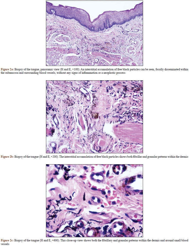
During his complete physical examination, a pigmented oral lesion was discovered: a black asymmetric 8-mm homogenous macule located at the right side of the tongue [Figure - 1]a. It was asymptomatic and the patient had not noticed it earlier. Dermoscopy imaging showed a structureless, homogeneous, black-bluish asymmetric macule with irregular but well-demarcated borders [Figure - 1]b. Biopsy revealed an interstitial accumulation of free black particles with both fibrillar and granular patterns, focally disseminated within the submucosa and surrounding blood vessels. No signs of inflammation or neoplastic processes were seen [Figure - 2].
 |
| Figure 1: (a) Black asymmetric homogenous macule on the right side of tongue. (b) Dermoscopic examination showing a structureless black-bluish asymmetric macule with irregular sharp borders |
 |
| Figure 2: |
What is Your Diagnosis?
| 1. |
Warszawik-Hendzel O, Slowinska M, Olszewska M, Rudnicka L. Melanoma of the oral cavity: Pathogenesis, dermoscopy, clinical features, staging and management. J Dermatol Case Rep 2014;8:60-6.
[Google Scholar]
|
| 2. |
Kauzman A, Pavone M, Blanas N, Bradley G. Pigmented lesions of the oral cavity: Review, differential diagnosis, and case presentations. J Can Dent Assoc 2004;70:682-3.
[Google Scholar]
|
| 3. |
Pigatto PD, Brambilla L, Guzzi G. Amalgam tattoo: A close-up view. J Eur Acad Dermatol Venereol 2006;20:1352-3.
[Google Scholar]
|
| 4. |
Olszewska M, Banka A, Gorska R, Warszawik O. Dermoscopy of pigmented oral lesions. J Dermatol Case Rep 2008;2:43-8.
[Google Scholar]
|
| 5. |
McCullough MJ, Tyas MJ. Local adverse effects of amalgam restorations. Int Dent J 2008;58:3-9.
[Google Scholar]
|
Fulltext Views
5,793
PDF downloads
4,146


