Translate this page into:
Primary amyloidosis
2 Department of Pathology, Apollo Hospitals, Jubilee Hills, Hyderabad, Andhra Pradesh, India
Correspondence Address:
Indukooru Subrayalu Reddy
A 8 Anand Sheel Enclave, Nandi Nagar, Road No. 14 Banjara Hills, Hyderabad - 500 033
India
| How to cite this article: Reddy IS, Swarnalata G. Primary amyloidosis. Indian J Dermatol Venereol Leprol 2016;82:178-179 |
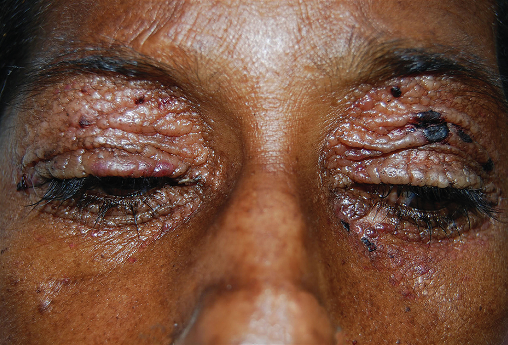
A 43-year-old woman with hypertension and chronic kidney disease on maintenance hemodialysis for 7 months developed multiple, asymptomatic, waxy papules and plaques, some of them hemorrhagic, over the eye lids, paranasal area [Figure - 1], neck, and lower abdomen since 4 months. She was admitted in the intensive medical care with altered sensorium, hypotension, gram-negative septicemia, severe anemia, and hypercalcemia. Skin biopsy of waxy papules showed atrophic epidermis and dense deposits of amyloid in the papillary dermis. Bone marrow aspiration revealed 31% plasma cells and serum protein immune electrophoresis showed a grossly elevated lambda free light chain level (1990 mg/dl). Correlating the clinical features, skin biopsy, and immune electrophoresis findings, she was diagnosed as having primary systemic amyloidosis secondary to multiple myeloma.
 |
| Figure 1: Waxy papules and plaques and hemorrhagic crusts involving both upper and lower eye lids |
Declaration of patient consent
The authors certify that they have obtained all appropriate patient consent forms. In the form the patient(s) has/have given his/her/their consent for his/her/their images and other clinical information to be reported in the journal. The patients understand that their names and initials will not be published and due efforts will be made to conceal their identity, but anonymity cannot be guaranteed.
Fulltext Views
3,051
PDF downloads
2,296


