Translate this page into:
Pseudo-lipomatosis cutis: A singular dermal artifact
Correspondence Address:
Rajiv Joshi
14 Jay Mahal, A Road, Churchgate, Mumbai - 400 020, Maharashtra
India
| How to cite this article: Joshi R. Pseudo-lipomatosis cutis: A singular dermal artifact. Indian J Dermatol Venereol Leprol 2015;81:504-505 |
Sir,
A 4 mm punch biopsy was received from a 33-year-old male with a severely itchy rash on the trunk and extremities of a few weeks duration, with nocturnal pruritus. The clinical differential diagnoses were prurigo and scabies.
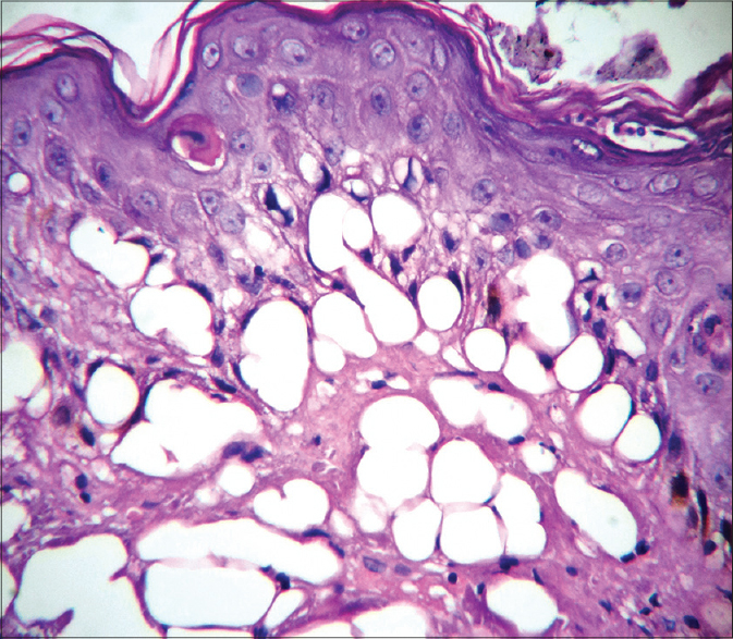
Histopathologic examination showed a hyperplastic epidermis with an occasional dyskeratotic keratinocyte in the upper spinous zone and a few neutrophils in the stratum corneum with flakes of parakeratosis. No scabetic mite was seen [Figure - 1].
 |
| Figure 1: Hyperplastic epidermis with a single dyskeratotic keratinocyte, few neutrophils in the stratum corneum and many vacuoles in the upper dermis. H and E, ×200 |
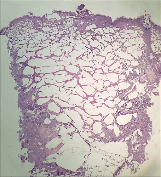
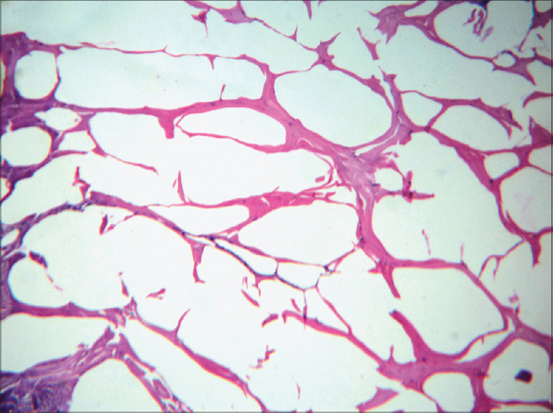
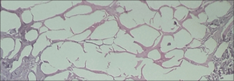
The entire dermis had innumerable, variably sized, empty spaces that resembled fat tissue and adipocytes without any inflammatory infiltrate. [Figure - 2] These spaces appeared to be located within the collagen of the dermis and were not lined by either endothelial or adipocyte nuclei [Figure - 3]. Normal adipose tissue was seen beneath the dermis riddled with the cystic spaces [Figure - 4].
 |
| Figure 2: The entire dermis is riddled with variably sized vacuoles and cystic spaces with poorly processed collagen at the periphery. H and E, ×40 |
 |
| Figure 3: Spaces within the dermal collagen without lining. H and E, ×100 |
 |
| Figure 4: Normal adipose tissue beneath the artifactually cystic dermis. H and E, ×40 |
The term pseudo-lipomatosis cutis [1] has been used to represent a likely artifactual change in the dermis with the appearance of numerous vacuoles and empty spaces that resemble the presence of fat tissue in the dermis. The visual appearance is very similar to mucosal pseudo-lipomatosis of the colon [2] where the variably sized empty spaces, which do not have any lining, are believed to represent accumulation of intestinal gas introduced at the time of endoscopy. Similarly, insufflation-induced gastric vacuolation has also been described. [3] Similar changes may occur in association with pneumatosis cystoides intestinalis.
True fatty infiltration of the dermis may occur in aging dermal melanocytic nevi, fibroepithelial polyps and in nevus lipomatosis superficialis as an atrophic or metaplastic phenomenon. [4] Dermal vacuoles may be seen within areas of dense dermal inflammation due to extracellular lipid deposition. In acrodermatitis chronica atrophicans, vacuoles in the upper dermis have been described in the setting of dermal sclerosis and have been attributed to lymphedema and the presence of prelymphatics that are walled off by ground substance and have no lining. [5]
Unlike the above situations, pseudo-lipomatosis cutis is an unusual dermal artifact unrelated to underlying pathologic processes and is postulated to occur due to injection of air during administration of local anesthetic prior to the biopsy procedure. Another proposed etiology for this artifact is the formation of gas in the tissue due to inadequate fixation with autolytic changes and gas bubble formation leading to empty spaces in the tissue. [6]
| 1. |
Trotter MJ, Crawford RI. Pseudolipomatosis Cutis: Superficial dermal vacuoles resembling fatty infiltration of the skin. Am J Dermatopathol 1998;20:443-7.
[Google Scholar]
|
| 2. |
Snover DC, Sandstad J, Hutton S. Mucosal pseudo-lipomatosis of the colon. Am J Clin Pathol 1985;84:575-80.
[Google Scholar]
|
| 3. |
Alper M, Akcan Y, Belenli OK, Cukur S, Aksoy KA, Suna M. Gastric pseudo-lipomatosis, usual or unusual? Re-evaluation of 909 endoscopic gastric biopsies. World J Gantroenterol 2003;9:2846-8.
[Google Scholar]
|
| 4. |
Maize JC, Foster G. Age-related changes in melanocytic naevi. Clin Exp Dermatol 1979;4:49-58.
[Google Scholar]
|
| 5. |
Brehmer-Andersson E, Hovmark A, Asbrink E. Acrodermatitis chronica atrophicans: Histopathologic findings and clinical correlations in 111 cases. Acta Derm Venereol 1998;78:207-13.
[Google Scholar]
|
| 6. |
Thomson SW, Luna LG. An atlas of artifacts encountered in the preparation of microscopic tissue sections. Springfield Illinois: Charles C. Thomas Publisher; 1978.p. 24.
[Google Scholar]
|
Fulltext Views
3,315
PDF downloads
1,574


