Translate this page into:
Sandpaper: An easy and effective tool to get rid of scales during the dermoscopic examination in plaque psoriasis
Corresponding author: Dr. Biswanath Behera, Department of Dermatology, and Venereology, All India Institute of Medical Sciences, Bhubaneswar, Odisha, India. biswanathbehera61@gmail.com
-
Received: ,
Accepted: ,
How to cite this article: Behera B, Dash S, Palit A. Sandpaper: An easy and effective tool to get rid of scales during the dermoscopic examination in plaque psoriasis. Indian J Dermatol Venereol Leprol 2021;87:599-600.
Problem
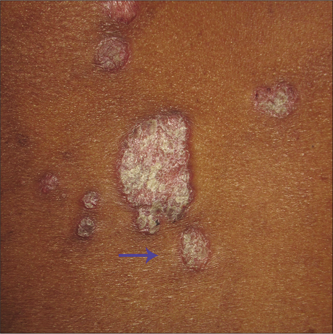
The presence of regularly distributed dotted vessels is the dermoscopic hallmark of plaque psoriasis. Golińska et al. reported the presence of the red dots/globules to be the most common vascular structure in psoriasis, the frequency of which ranged from 97.1% to 100%.1 The regular pattern of distribution of dotted vessels enables one to distinguish psoriasis from its clinical mimics. A case of clear cell acanthoma within a psoriatic plaque that demonstrated dotted vessels in a linear pearl-like distribution could be recognized due to the difference in the pattern of distribution between the two conditions.2 This highlights the importance of dotted vessels for the diagnosis of plaque psoriasis. The silvery-white scales on the plaque pose a significant problem in demonstrating this dermoscopic feature [Figure 1a].

- Psoriatic plaque (arrow) before the use of sandpaper
Solution
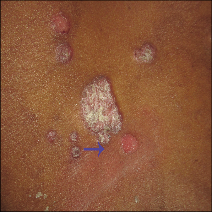
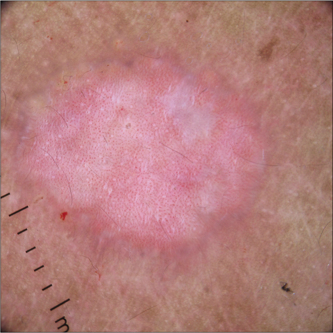
To overcome this difficulty, sandpaper was used to remove the scales. The scales were easily and effectively removed without any significant pain to the patient. During the procedure, all the scales fell off along with the Bulkeley’s membrane [Figure 1b]. This enabled us to visualize the regularly distributed dotted vessels under dermoscope [Figure 1c], without any significant bleeding, as demonstrated in Video 1. The conventional use of glass slide removes the scales in layers followed by removal of Bulkeley’s membrane, that allows the demonstration of underlying dotted vessels; it can be painful and the sharp edge of the glass slide may cause bleeding. As per dermoscopy of inflammatory conditions is concerned, scale plays a significant role in the derivation of accurate diagnosis. They are bright white, dull-white, focal and collarette in psoriasis, lichen planus, eczema and pityriasis rosea, respectively. The examination of scales, both its color and distribution, is also important to arrive at a diagnosis. However, scales should be removed when hyperkeratotic lesions are present on the palms and soles or to visualize underlying dermoscopic features.

- Psoriatic plaque (arrow) after the use of sandpaper, showing complete absence of the scales without bleeding points

- Dermoscopic image of the plaque demonstrating regularly distributed dotted vessels on a pinkish background (Dermlite DL4, ×10)
Declaration of patient consent
The authors certify that they have obtained all appropriate patient consent.
Financial support and sponsorship
Nil.
Conflicts of interest
There are no conflicts of interest.
References
- Dermoscopic features of psoriasis of the skin, scalp and nails-A systematic review. J Eur Acad Dermatol Venereol. 2019;33:648-60.
- [CrossRef] [PubMed] [Google Scholar]
- Dermoscopy, “clears” out the diagnosis: An erythematous nodule in a psoriatic patient. J Am Acad Dermatol. 2015;72:S68-70.
- [CrossRef] [PubMed] [Google Scholar]


