Translate this page into:
Schamberg's disease - an unusual presentation
Correspondence Address:
S R Sengupta
S A, Binoy Bala Mukherjee Lane, Uttarpara, W. Bengal - 712258,
India
| How to cite this article: Sengupta S R, Malakar S, Lahiri K. Schamberg's disease - an unusual presentation. Indian J Dermatol Venereol Leprol 1997;63:189-190 |
Abstract
A 28-year-old man with typical lesions of Schamberg's progressive pigmented purpuric dermatosis involving his right forearm is reported here for its unusual localisation. |
 |
 |
 |
Schamberg′s disease denotes a progressive pigmentary dermatois involving usually the lower part of the legs. Irregular patches of orange or brown pigmentation due to hemosiderin, with characteristic ′cayanne paper′ spots. [1,2] The lesions are chronic and may be pruritic.
It is one of the constituent of the entity purpura pigmentosa chronica, others being purpura annularis telangiectodes of Majocchi, pigmented purpuric lichenoid dermatitis of Gougerot and Blum, and eczematid-like purpura of Doucas and Kaperanakis.[2] They are so closely related to each other, both clinically and histologically, that differentiation becomes very difficult and also unnecessary.[3] Purpura pigmentosa chronica appears to be suitable term to denote these diseases.
Case Report
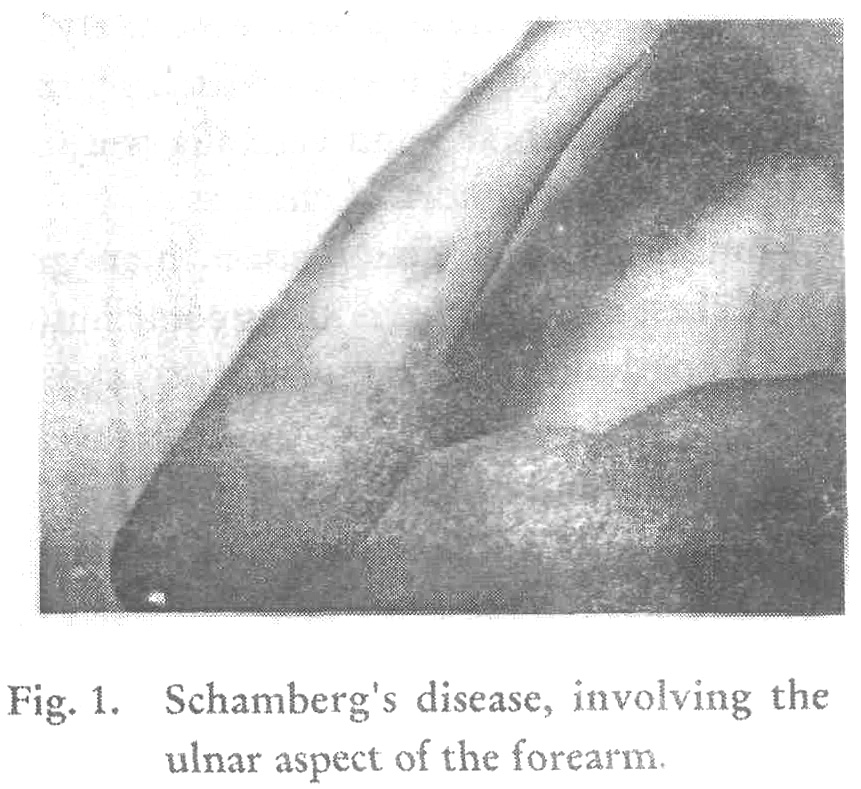
A 28-year-old male patient reported at our OPD with pigmented patches over his right forearm. On examination, the lesions were found to be brown in colour, irregularly shaped with typical ′cayenne paper′ spots in the centre and over the edges [Figure - 1]. They were distributed more along the ulnar aspect of the forearam. Small new macules were also present just above the right elbow. The lesions were present for nearly a year and were progressing slowly. Other than slight pruritus they were symptomless.
Other parts of his body, including his left hand and both the legs were absolutely free from such type of lesions. His hair, nails, mucous membrane and genitalia were also free from any abnormal changes.
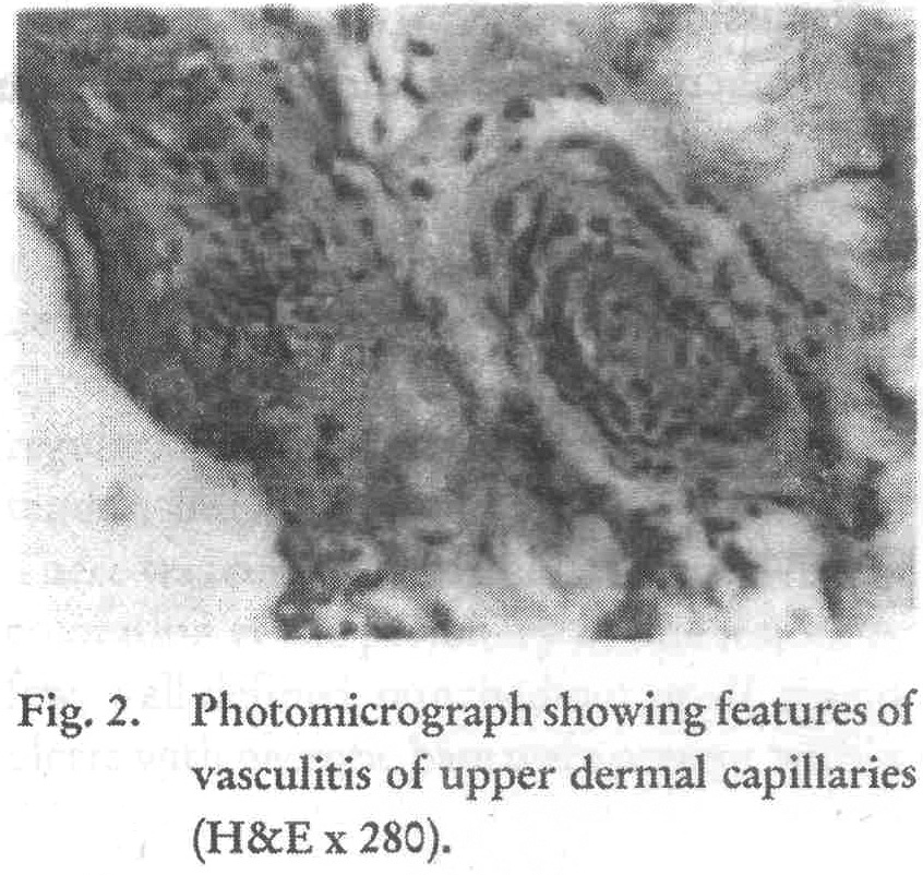
Routine physical examination, hemogram and chest x-ray yielded nothing contributory. Histopathological examination of skin biopsy specimen from a typical lesion revealed dialatation and swelling of upper dermal capillaries and a cellular infiltrate, consisting largely of lymphocytes arranged especially near the capillaries, thus giving a picture of lymphocytic type of vasculitis. [Figure - 2].
Discussion
Schamberg′s disease is known to occur usually over the lower limbs but rarely generalised eruption may occur. Involvement of the hands are very rare. Moyer reported a rare case of capillaritis of the palms.[4] In our case the clinical diagnosis of Schamberg′s disease was made provisionally and was subsequently proved histopathologically. But the unusual localisation of the disease in the upper extremity and the peculiar unilateral distribution of the lesions have made this case an unique one.
| 1. |
Champion R H. Purpura, in: Text Book of Dermatology, (Champion R H, Burton J L, Ebling F J G), 5th edn, Blackwell Scientific Publication, London Oxford 1992;1881-1892.
[Google Scholar]
|
| 2. |
Lever W F, Lever G S Histopathology of Skin, 7th edition, JB Lippincott company, Philadelphia, 1990;185-209.
[Google Scholar]
|
| 3. |
Randall S J, Kierland R R, Montgomery H. Pigmented purpuric eruptions, Arch Dermatol Syphilol 1951;64:177-191.
[Google Scholar]
|
| 4. |
Moyer D G, Chernita S A. Capillaritis of the palms, Arch Dermatol, 1969;99:591-592.
[Google Scholar]
|


