Translate this page into:
Serum galectin-9 levels in atopic dermatitis, psoriasis and allergic contact dermatitis: A cross-sectional study
2 Department of Clinical Pathology, Faculty of Medicine, Zagazig University, Zagazig, Egypt
Correspondence Address:
Eman Nofal
Department of Dermatology and Venereology, Faculty of Medicine, Zagazig University, Zagazig
Egypt
| How to cite this article: Nofal E, Eldesoky F, Nofal A, Abdelshafy A, Zedan A. Serum galectin-9 levels in atopic dermatitis, psoriasis and allergic contact dermatitis: A cross-sectional study. Indian J Dermatol Venereol Leprol 2019;85:195-196 |
Sir,
Galectin-9 (Gal-9) is a tandem-repeat galectin that is widely distributed in human tissues, including epithelial tissues. Galectin-9 is a physiological ligand for T-cell-immunoglobulin- and mucin-domain-containing molecule-3 (TIM-3) that is specifically expressed in T helper 1 cells (Th1) and T helper 17 cells (Th17), but not in T helper 2 cells (Th2). The interaction of TIM-3 and Gal-9 induces apoptosis in Th1 and Th17 cells.[1] Dysregulation of Gal-9-TIM-3 network probably causes several allergic or autoimmune diseases.[2] Atopic dermatitis, psoriasis and allergic contact dermatitis are common inflammatory skin diseases associated with immunological abnormalities. Atopic dermatitis is characterized by Th2 cell predominance, which is proposed to play a key pathogenetic role in atopic dermatitis, while psoriasis and allergic contact dermatitis are generally thought to be mediated by Th1 and Th17 cells. The aim of this work was to investigate a potential role of Gal-9 in the pathogenesis of atopic dermatitis, psoriasis and allergic contact dermatitis and to assess its relation to disease severity.
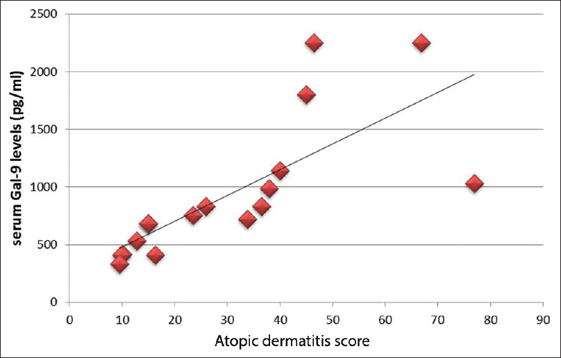
A total of 54 patients (18 atopic dermatitis, 18 psoriasis, 18 allergic contact dermatitis) with different grades of disease severity and 18 healthy subjects were recruited at the outpatient clinics of dermatology and venereology department, Zagazig University Hospitals. The severity of the diseases was determined by SCORAD in atopic dermatitis cases, by PASI score in psoriasis cases and by staging of allergic contact dermatitis. Serum Gal-9 levels were measured in all patients and controls by enzyme-linked immunosorbent assay. The mean serum levels of Gal-9 were 291.89 ± 67.66 pg/ml in control group, 1199 ± 697.65 pg/ml in atopic dermatitis group, 332.98 ± 61.3 pg/ml in psoriasis and 325.6 ± 64.67 pg/ml in allergic contact dermatitis group [Figure - 1]. A highly significant elevation in serum levels of Gal-9 in atopic dermatitis patients was detected (P = 0.001) and was positively correlated with the disease severity (P = 0.03). The more the severity of atopic dermatitis, the higher the serum levels of Gal-9 [Figure - 2]. No statistical differences were detected between the serum levels of Gal-9 in psoriasis and allergic contact dermatitis groups and the control group (P = 0.07 and 0.12, respectively). Moreover, there were no significant associations between the severity of psoriasis or allergic contact dermatitis and serum levels of Gal-9 (P = 0.15 and 0.49, respectively).
 |
| Figure 1: Mean serum galectin-9 levels among the three studied groups and controls |
 |
| Figure 2: Positive correlation between the severity of atopic dermatitis and serum galectin-9 levels |
As far as we know, the study of Nakajima et al. is the only study that investigated serum Gal-9 in atopic dermatitis and reported elevated serum Gal-9 levels in patients than controls and showed stronger expression of Gal-9 on epidermal keratinocytes and mast cells in atopic dermatitis skin than in normal skin.[3] They also found a significant decrease in the serum levels of Gal-9 with improvement of the skin lesions of atopic dermatitis after treatment. Several mechanisms have been suggested to explain the role of Gal-9 in atopic dermatitis. Gal-9 induces apoptosis in Th1 and Th17 cells through binding with its ligand TIM-3 resulting in Th2 polarization. The Th2 polarized microenvironment in lesional skin of atopic dermatitis induces Gal-9 upregulation with subsequent exacerbation of Th2 polarization. Dendritic cellsplay a crucial role in Th2 skewing, and mediate the development of atopic dermatitis. The role of Gal-9 in dendritic cell maturation has been assessed. The role of eosinophils in the pathogenesis of atopic dermatitis has been suggested. Gal-9 has an eosinophil attractant effect. Increased levels of Gal-9 in atopic dermatitis may have a role in eosinophilia detected in atopic patients. Increased mast cell numbers and activation were reported in chronic skin lesions of atopic dermatitis. The role of mast cells in the pathogenesis of atopic dermatitis is controversial. Gal-9 has a dual role in the function of mast cells.
In the psoriatic group, our results supported those of de la Fuente et al. who reported that the expression of Gal-9 in psoriatic skin and control skin was the same. However, they did not measure its levels in serum. We could not find previous studies investigating Gal-9 expression (either in serum or tissue) in allergic contact dermatitis. Downregulation of Gal-9 level was expected in cases of psoriasis and allergic contact dermatitis which could promote the Th1 and Th17 immune response observed in psoriasis and allergic contact dermatitis. Although this study may suggest that Gal-9 has no role in the pathogenesis of either disease, the study of Niwa et al. reported marked therapeutic effect in induced psoriasis and allergic contact dermatitis in mouse models by administration of stable Gal-9 and suggested Gal-9 as a unique and useful therapeutic tool for the treatment of Th1 and/or Th17 mediated skin disorders.[5] In conclusion, Gal-9 may be implicated in the pathogenesis of atopic dermatitis and is correlated to disease severity. Its role in psoriasis and allergic contact dermatitis could not be verified by this study. Restoration of the immune equilibrium lost in atopic dermatitis, psoriasis and allergic contact dermatitis by galectins require increased recognition of these molecules as immunoregulatory molecules and as a new therapeutic tool.
Declaration of patient consent
The authors certify that they have obtained all appropriate patient consent forms. In the form the patients have given their consent for their images and other clinical information to be reported in the journal. The patients understand that their names and initials will not be published and due efforts will be made to conceal their identity, but anonymity cannot be guaranteed.
Financial support and sponsorship
Nil.
Conflicts of interest
There are no conflicts of interest.
| 1. |
Kashio Y, Nakamura K, Abedin MJ, Seki M, Nishi N, Yoshida N, et al. Galectin-9 induces apoptosis through the calcium-calpain-caspase-1 pathway. J Immunol 2003;170:3631-6.
[Google Scholar]
|
| 2. |
Anderson AC, Anderson DE. TIM-3 in autoimmunity. CurrOpin Immunol 2006;18:665-9.
[Google Scholar]
|
| 3. |
Nakajima R, Miyagaki T, Oka T, Nakao M, Kawaguchi M, Suga H, et al. Elevated serum galectin-9 levels in patients with atopic dermatitis. J Dermatol 2015;42:723-6.
[Google Scholar]
|
| 4. |
de la Fuente H, Perez-Gala S, Bonay P, Cruz-Adalia A, Cibrian D, Sanchez-Cuellar S, et al. Psoriasis in humans is associated with down-regulation of galectins in dendritic cells. J Pathol2012;228:193-203.
[Google Scholar]
|
| 5. |
Niwa H, Satoh T, Matsushima Y, Hosoya K, Saeki K, Niki T, et al. Stable form of galectin-9, a Tim-3 ligand, inhibits contact hypersensitivity and psoriatic reactions: A potent therapeutic tool for Th1 – and/or Th17-mediated skin inflammation. Clin Immunol 2009;132:184-94.
[Google Scholar]
|
Fulltext Views
3,514
PDF downloads
1,755


