Translate this page into:
Skin discoloration: A new sign of systemic candidiasis in neonates?
Correspondence Address:
B Vishnu Bhat
Neonatology Division, Department of Pediatrics, JIPMER, Pondicherry 605 006
India
| How to cite this article: Femitha P, Joy R, Prasad K, Gane BD, Adhisivam B, Bhat B V. Skin discoloration: A new sign of systemic candidiasis in neonates?. Indian J Dermatol Venereol Leprol 2012;78:408 |
Sir,
With advances in neonatal care, there is increased survival of very low birth weight (VLBW) infants. The use of invasive procedures like endotracheal intubation, placement of central intravenous lines and invasive monitoring techniques, combined with risks of prolonged ICU stay and need for broad-spectrum antibiotics has led to the emergence of new and resistant pathogens including fungi. Fungal septicemia is the third most common cause of late onset sepsis. Its incidence varies with many factors, but generally it is inversely proportional to the gestational maturity and birth weight. An overall incidence of 1-4% and mortality as high as 25-50% has been reported. [1] Various muco-cutaneous manifestations of fungal septicemia have been described in literature.
Cutaneous involvement occurs in 50-60% of VLBW neonates with systemic candidiasis. Congenital cutaneous candidiasis is a distinct entity first described by Benirschke and Raphael in 1958. The extensive rash, usually appears within 12 hours of birth. [2] It presents as an erythematous, maculopapular eruption of the trunk and extremities which rapidly becomes pustular, then improves with generalized desquamation. Most often, it resolves spontaneously in term infants, but may lead to invasive disease and result in mortality in VLBW babies. Colonization of skin and gastrointestinal tract (GIT) with Candida sp. forms an important and initial step in the pathogenesis of systemic infection. [3] Kaufman et al., reported that the skin and GIT are colonized before the respiratory tract. Involvement of the skin in acquired infection may present as erythematous, scaling dermatitis with satellite papules and pustules. [4] Intertriginous infection usually involving the groin may be associated with oral thrush. Another rare manifestation reported by Baley and Silverman resembled widespread first-degree burns whose desquamation resulted in superficial erosions. [5] In 1991, ulcerative and erosive lesions with tightly adherent white crusts were described in extremely low birth weight neonates and their skin biopsies showed invasion from the epidermis to the dermis. [2]
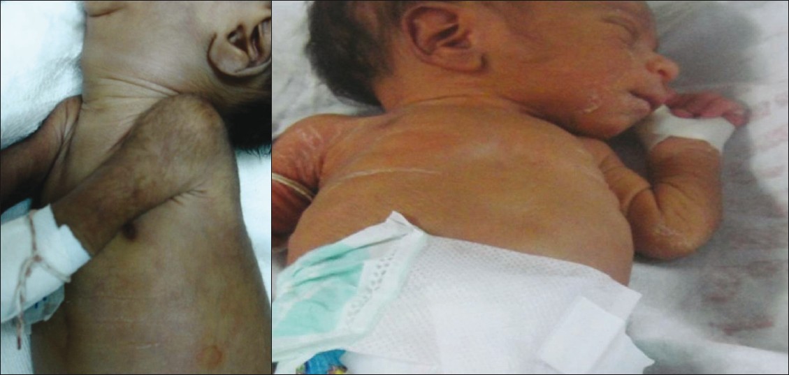
We report a generalized grayish brown skin color change in neonates with fungal sepsis [Figure - 1]a. This finding was noted in 25 out of 36 cases of blood culture proven candidal infection during the period from October 2009 to June 2011. All the babies in the study group were from the same ethnic population of South India. Of the 36 neonates who developed candidal sepsis, 28 were preterm and 8 were term. Twenty three of the 25 who developed skin color change were preterm and 2 babies were term. There were no significant differences in the morbidity and mortality of neonates who developed the described color change compared to those who did not.
 |
| Figure 1a: Skin color change - generalized grayish brown discoloration Figure 1b: Normal skin color after 3 - 4 days of antifungal therapy |
Although many of these babies with fungal sepsis had known manifestations like bleeding, feed intolerance and hyperglycemia, they were also noted to have concomitant discoloration of skin all over the body. This was in the absence of hypothermia, systemic hypoperfusion/shock or jaundice which could explain color change. At the time of noticing color change, none of the babies had received phototherapy. Blood culture done in 25 neonates revealed Candida albicans in 7(28%), C. glabrata in 11(44%), C. parapsilosis in 6(24%) and C. tropicalis in 1(4%). The overall mortality was 44.4%. It was also interesting to note that the color improved and almost returned to normal after institution of either azole or amphotericin B [Figure - 1]b. Normal color returned after an average duration of 3.2 ± 2.14 days of antifungal therapy. This change could possibly be due to vasomotor instability or due to the candidemia per se invading blood vessel walls. We could not do a skin biopsy as most of these babies were preterm and sick. This skin change has not been previously reported and hitherto unique to our cases.
Many studies in the past have emphasized the need for early and empirical antifungal therapy in those suspected to have infection to improve survival and reduce morbidity. Our observation may improve the case detection rates and thus have implications on commencing early antifungal therapy.
| 1. |
Saiman L, Ludington E, Pfaller M, Rangel-Frausto S, Wiblin RT, Dawson J, et al. Risk factors for candidemia in neonatal intensive care unit patients. The national epidemiology of mycoses survey study group. Pediatr Infect Dis J 2000;19:319-24.
[Google Scholar]
|
| 2. |
Rowen JL, Atkins JT, Levy ML, Baer SC, Baker CJ. Invasive fungal dermatitis in the ≤1000 g neonate. Pediatrics 1995;95:682-7.
[Google Scholar]
|
| 3. |
Baley JE, Kliegman RM, Boxerbaum B, Fanaroff AA. Fungal colonization in the very low birth weight infant. Pediatrics 1986;78:225-32.
[Google Scholar]
|
| 4. |
Kaufman DA, Gurka MJ, Hazen KC, Boyle R, Robinson M, Grossman LB. Patterns of fungal colonization in preterm infants weighing <1000 g at birth. Pediatr Infect Dis J 2006;25:733-7.
[Google Scholar]
|
| 5. |
Baley JE, Silverman RA. Systemic candidiasis: Cutaneous manifestations in low birth weight infants. Pediatrics 1988;82:211-5.
[Google Scholar]
|
Fulltext Views
2,426
PDF downloads
1,880


