Translate this page into:
Solitary tumor with a peripheral depigmented patch and poliosis
Correspondence Address:
Chien-Ping Chiang
Department of Dermatology Tri-Service General Hospital No. 325, Sec. 2, Chenggong Rd., Neihu Dist., Taipei City 114
Taiwan
| How to cite this article: Hung CT, Chiang CP. Solitary tumor with a peripheral depigmented patch and poliosis. Indian J Dermatol Venereol Leprol 2012;78:776 |
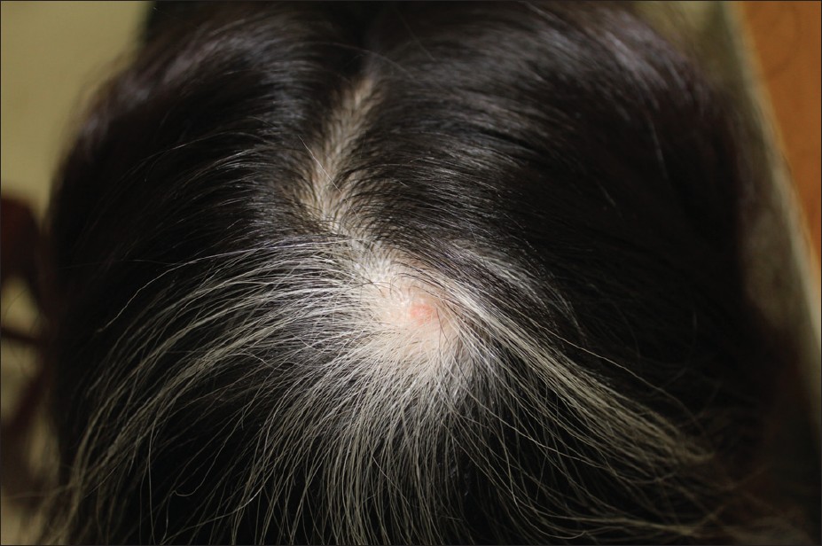
An 11-year-old Taiwanese girl presented with a 2-year history of a flesh-colored tumor over a patch of white hair on the vertex of the scalp. Initially, the tumor was hyperpigmented, without any surrounding depigmented areas or white hairs since preschool age. Her medical history was unremarkable, and mental and developmental milestones were normal.
Dermatological examination revealed a 0.8-cm diameter, flesh-colored tumor surrounded by a 2-cm diameter, well-circumscribed depigmented halo with poliosis [Figure - 1]. There were no other white patches visible under a Wood′s lamp examination.
 |
| Figure 1: A 0.8 cm diameter, flesh-colored tumor surrounded by a 2 cm diameter, circumscribed depigmented halo with poliosis |
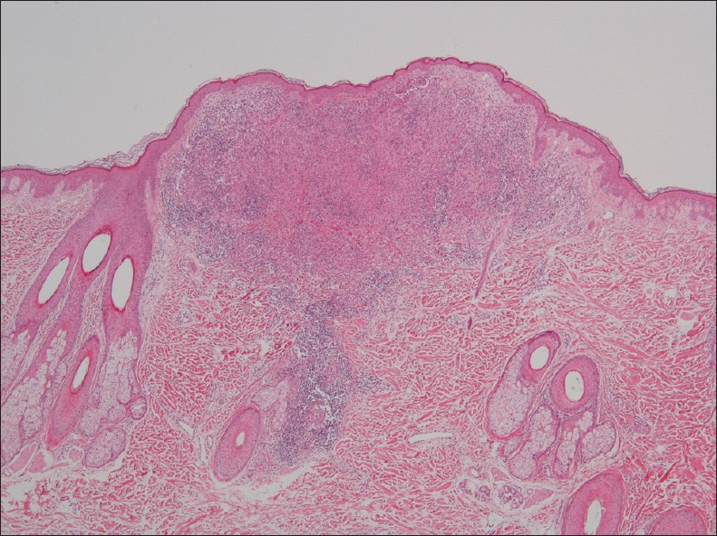
Histopathological examination of a skin biopsy taken from the scalp tumor showed several nests of pale-staining epithelioid cells with moderately dense infiltration by lymphocytes and multiple giant cells in the papillary dermis [Figure - 2] and [Figure - 3]. The epithelioid cells were positive for the S-100 protein and melan-A [inset in [Figure - 3]].
 |
| Figure 2: Histopathology showing a wedge-shaped infiltration with nests of pale-staining epithelioid cells surrounded by dense lymphocytes in the papillary dermis (H and E, ×40) |
 |
| Figure 3: Histopathology showing multiple giant cells with wreath-like nuclei in the papillary dermis (H and E, ×400); inset showing positive melan-A staining in these cells (×400) |
What′s Your Diagnosis?
Diagnosis
Halo nevus associated with poliosis circumscripta
Discussion
Halo nevus, also known as leukoderma acquisitum centrifugum, is a melanocytic nevus surrounded by a depigmented ring. It shows a predilection for the back and mostly affects children and young adults, with a mean age of 16 years. [1] Histopathologically, it is characterized by the presence of nevomelanocytic cells surrounded by mixed inflammatory cells, including lymphocytes, histiocytes, and occasional mast and plasma cells. Epithelioid granuloma may sometimes be present. There are several other cutaneous tumors that exhibit the clinical halo phenomenon, including neurofibroma, histiocytoma, basal cell carcinoma, sarcoidosis, and lichen planus. It is necessary to combine clinical and pathological findings for the correct diagnosis. Poliosis is defined as localized white hairs and is associated with several lesions, including congenital nevus, neurofibroma, intradermal nevus, and melanoma. This condition is a rare presentation of halo nevus.
There were several reports of halo nevus associated with the development of vitiligo. [2],[3] Gutte et al[3] reported a 22-year-old man who sustained a halo nevus with poliosis over his occipital scalp and developed extralesional vitiligo over his left knee. The authors suggested that halo nevus should be excised as early as possible to prevent progression to vitiligo. [3] We conducted a Wood′s lamp examination on our patient, but did not find any other whitish patches or halo nevi in her body.
The mechanism underlying the development of halo nevus is still uncertain. On the basis of the infiltration of mononuclear cells observed in most halo nevi, it is possible that an immunologic response plays a role in the pathogenesis of the condition. Zeff et al[4] proposed that cytotoxic CD8 + T cells are the effectors that destroy nevomelanocytes. Gutte et al[3] proposed that CD8 + T cells may also be related to the vitiligo lesions located distant to the nevi.
The differential diagnosis of the present case includes juvenile xanthogranuloma, which is a benign histiocytic disorder with a predilection for the head and neck that principally appears in the first 2 decades of life. Histopathological features include the presence of monomorphous histiocytes and Touton giant cells with a ring of nuclei surrounded by a foamy cytoplasm. [5] In our patient, we observed numerous giant cells with wreath-like nuclei, but no vacuolated cytoplasm. Immunohistochemistry was helpful for distinguishing nevomelanocytic cells from histiocytes. On the basis of the positive staining for S-100 protein and melan-A, we confirmed that the epithelioid cells were, in fact, nevomelanocytic cells.
When clinical dermatologists find patients with poliosis, the diagnosis of halo nevus should be considered. Due to the association between halo nevi and vitiligo, we suggest a thorough Wood′s lamp examination.
| 1. |
Mooney MA, Barr RJ, Buxton MG. Halo nevus or halo phenomenon? A study of 142 cases. J Cutan Pathol 1995;22:342- 8.
[Google Scholar]
|
| 2. |
Hofmann UB, Brocker EB, Hamm H. Simultaneous onset of segmental vitiligo and a halo surrounding a congenital melanocytic naevus. Acta Derm Venereol 2009;89:402-6.
[Google Scholar]
|
| 3. |
Gutte R, Khopkar U. Extralesional vitiligo over knee following halo nevus with poliosis on the scalp. Indian J Dermatol Venereol Leprol 2011;77:522-5.
[Google Scholar]
|
| 4. |
Zeff RA, Freitag A, Grin CM, Grant-Kels JM. The immune response in halo nevi. J Am Acad Dermatol 1997;37:620-4.
[Google Scholar]
|
| 5. |
Dehner LP. Juvenile xanthogranulomas in the first two decades of life: A clinicopathologic study of 174 cases with cutaneous and extracutaneous manifestations. Am J Surg Pathol 2003;27:579-93.
[Google Scholar]
|
Fulltext Views
5,694
PDF downloads
3,031


