Translate this page into:
Sporotrichoid pattern of malignant melanoma
Correspondence Address:
Ranjan C Rawal
2, Friends Avenue, S.G. Road, Ahmedabad
India
| How to cite this article: Rawal RC, Mangla K. Sporotrichoid pattern of malignant melanoma. Indian J Dermatol Venereol Leprol 2008;74:70-71 |
 |
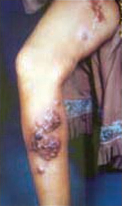
| Figure 1: Multiple deep seated nodules of malignant melanoma in sporotrichoid pattern |
 |
| Figure 1: Multiple deep seated nodules of malignant melanoma in sporotrichoid pattern |
Sir,
Malignant melanoma is a highly invasive neoplasm of skin, strongly influenced by environmental factors that develop in a genetically susceptible host. Incidence of melanoma is rising in Caucasians although it is a rare presentation in India. [1]
A 28-year-old female presented with multiple painful lesions over her right leg of eight months duration. She was a farmer and claimed that the lesions started as a single painful nodule on the heel following a thorn prick. She ignored it then and subsequently the size and number of the lesions increased to involve the whole right lower limb as linearly arranged nodules. Cutaneous examination revealed multiple tender nodulo-ulcerative lesions with discharge, arranged in linear fashion from heel to thigh, present over the right leg [Figure - 1]. Other findings included edema of the feet, thickened lymphatic channels between nodules along with bilateral stony hard, tender inguinal lymph nodes. The patient was previously diagnosed as sporotrichosis and treated with antifungal (itraconazole) drugs for six months in another healthcare facility without any clinical response. A differential diagnosis of sporotrichosis and malignant melanoma was considered. Limb X-ray, KOH smear, gram stained smear and fungal culture were negative. Skin biopsy revealed atypical melanocytes in the epidermis and dermis in nests. General examination revealed multiple generalized lymphadenopathy (axillary, cervical and submandibular lymph nodes). Abdominal sonography showed liver metastasis. Chest X-ray was normal. A diagnosis of malignant melanoma was established and the patient was referred to the oncology department for further management.
The incidence of melanoma continues to rise at an epidemic rate as evidenced by a 101.5% increase from the 1970s to the 1990s. [2],[3] Melanoma represents the fifth most common type of cancer, the most common type in women 25-29 years of age and the most common type in Caucasian men 25-44 years of age. But it is rare in Indian patients. Nodular melanoma and melanoma d′emblee are rare types of primary cutaneous malignant melanoma that are invasive and lack intraepidermal component. [4] These lesions when first noted clinically are always palpable, convex in shape, of varying shades, rapidly increasing in size; neglected tumors may be several centimeters in diameter. Ulceration occurs fairly early. They can occur in any portion of the skin/mucosa. [4]
Our patient presented with history of trauma with multiple nodular ulcerative lesions arranged in a linear fashion along the lymphatics over the lower limb which clinically simulated lymphocutaneous sporotrichosis. However, histopathology helped us to reach the correct diagnosis.
| 1. |
Johnson TM, Chang A, Redman B, Rees R, Bradford C, Riba M, et al. Management of Melanoma with a multi disciplinary melanoma clinic model. J Am Acad Dermatol 2000;42:820-6.
[Google Scholar]
|
| 2. |
Rigel DS, Friedman RJ, Kopf AW. The incidence of malignant melanoma in United States: Issues as we approach the 21 st century. J Am Acad Dermatol 1996;34:839-47.
[Google Scholar]
|
| 3. |
Chang AE, Karmel LH, Menck HR. The national cancer data base report on cutaneous and non cutaneous melanoma: A summary of 84, 836 cases from the past decades. Cancer 1998;8:1664-78.
[Google Scholar]
|
| 4. |
McGovern VJ, Mihm MC, Baily C, Booth JC, Clark WH, Cochran AJ. The classification of malignant melanoma and its histological reporting. Cancer 1973;32:1446-57.
[Google Scholar]
|
Fulltext Views
2,829
PDF downloads
2,618


