Translate this page into:
Quiz
2007:73:6;447-448
doi: 10.4103/0378-6323.37084
PMID: 18032880
Scaly erythematous plaque on the chest
Vandana Mehta Rai1 , C Balachandran1 , Sudha Bhat2 , V Geetha2
1 Department of Skin and STD, KMC, Manipal, India
2 Department of Pathology, KMC, Manipal, India
Correspondence Address:
Vandana Mehta Rai
Department of Skin and STD, Kasturba Medical College, Manipal - 576 104, Karnataka
India
How to cite this article:
Rai VM, Balachandran C, Bhat S, Geetha V. Scaly erythematous plaque on the chest. Indian J Dermatol Venereol Leprol 2007;73:447-448 |
Copyright: (C)2007 Indian Journal of Dermatology, Venereology, and Leprology
|
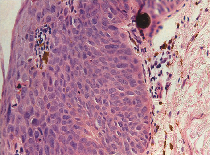
Figure 4: Skin biopsy (H and E stain, X400) |
|
Figure 4: Skin biopsy (H and E stain, X400) |
|
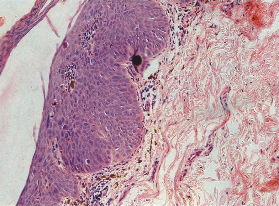
Figure 3: Skin biopsy (H and E stain, X200) |
|
Figure 3: Skin biopsy (H and E stain, X200) |
|
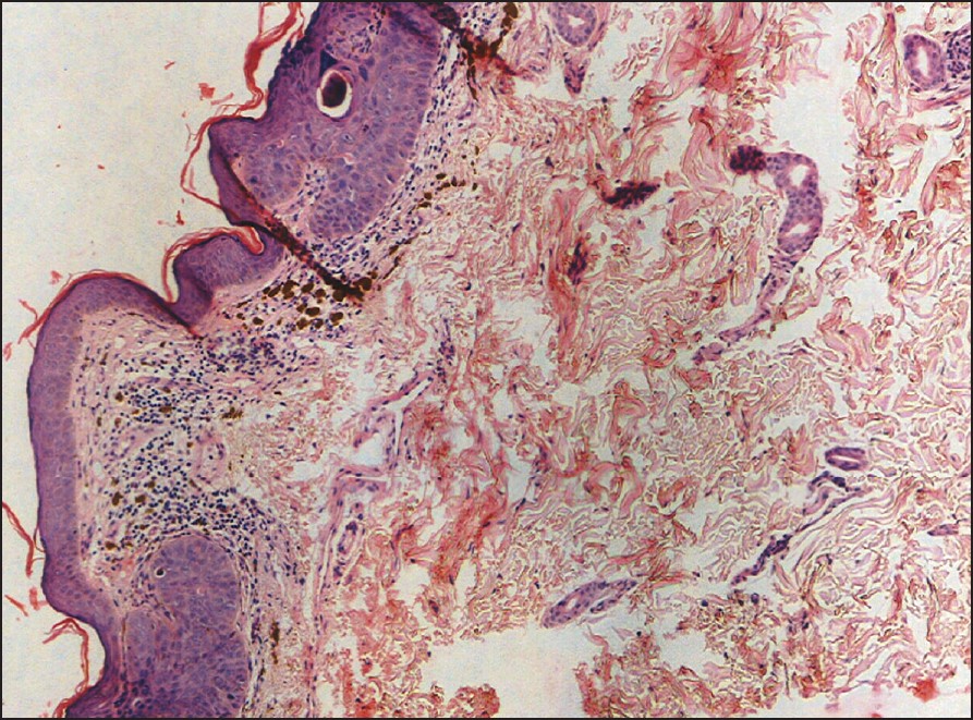
Figure 2: Skin biopsy (H and E stain, X100) |
|
Figure 2: Skin biopsy (H and E stain, X100) |
|
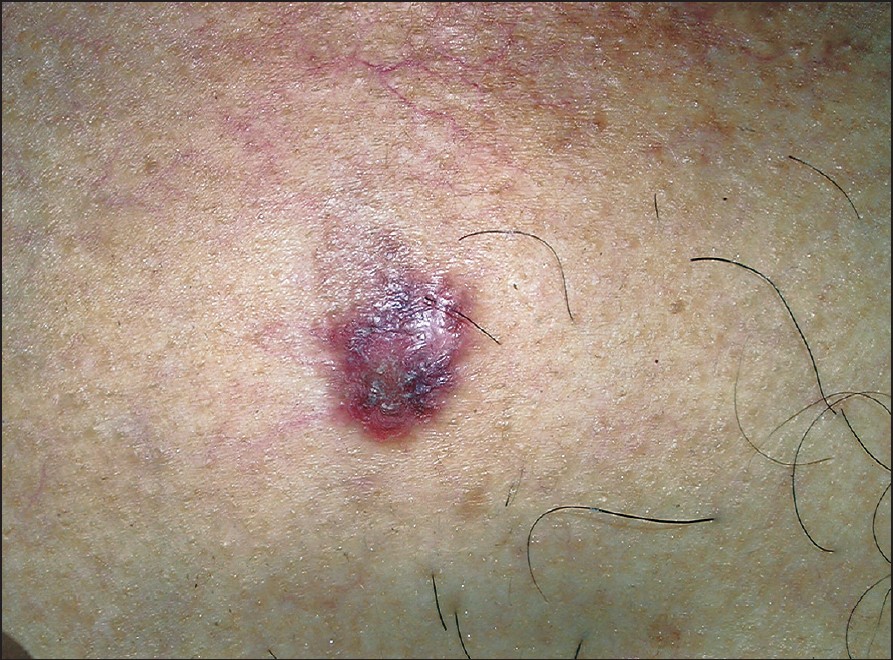
Figure 1: Scaly erythematous plaque on the chest |
|
Figure 1: Scaly erythematous plaque on the chest |
A 55 year-old male presented to us with an asymptomatic plaque on the chest of a year′s duration. There was a history of excessive exposure to the sun as he was a field worker but there was no history of contact with insecticides, pesticides or fertilizers. There was no similar lesion elsewhere on the body. The patient had been treated by a local physician with topical steroids for the past one year without any improvement. Cutaneous examination revealed a 3 x 3 cm well-marginated, erythematous, mildly scaly plaque on the V area of the neck [Figure - 1]. Biopsy performed from the lesion showed an irregularly acanthotic epidermis with mild nuclear pleomorphism, loss of normal architecture, scattered dyskeratotic keratinocytes, multinucleated giant cells with a lichenoid lymphoplasmacytic infiltrate [Figure - 2],[Figure - 3],[Figure - 4].
What is your Diagnosis ?
View Answer
Diagnosis: Bowen′s disease
Discusson
Bowen′s disease (BD) represents a form of squamous cell carcinoma in situ that appears as a slowly enlarging, sharply marginated, erythematous, scaly or crusted plaque. It may occur on both exposed and unexposed skin. Chronic sun exposure plays an important role in its development as lesions predominantly occur on the sun-exposed areas of the head and the neck. In addition, involvement of the lower limbs below the knees further supports the role of sun exposure in the pathogenesis of BD. [1] When BD occurs on unexposed areas, a history of arsenic intake should be excluded as long-term arsenic exposure has also been implicated in the development of BD. Agricultural workers are particularly at risk as they frequently take inadequate precautions against accidental ingestion or inhalation of arsenic-containing insecticides or pesticides. [2] Of late, human papilloma virus (HPV) has been implicated in the development of BD and HPV 2, 16, 34 and 35 have been detected recently in 20 cases of nongenital BD. [3] The lesion presents as an asymptomatic, well demarcated, erythematous and a mildly scaly plaque which enlarges gradually. Development of ulceration is usually a sign of invasive carcinoma and may be delayed for many years. When there is good evidence of chronic arsenicalism either by history or because of associated signs of arsenic keratoses and mottled pigmentation, the possibility of a visceral malignancy especially of the lung should be borne in mind. Less easily recognised variants of BD are pigmented, verrucous, fungating and BD of the nail bed. Histopathologically, BD is characterised by an acanthotic epidermis with elongation and thickening of rete ridges. Throughout the epidermis, the cells lie in complete disorder resulting in a ′windblown appearance′. Many cells appear highly atypical showing large hyperchromatic nuclei. Another common characteristic feature is the presence of cells showing atypical, individual cell keratinisation. Even though there is marked atypicality of the epidermal cells, the border between the epidermis and dermis appears sharp and the basement membrane remains intact. The upper dermis shows a moderate amount of chronic inflammatory infiltrate.
Treatment options for BD include surgical excision, cryotherapy, curettage, cautery, chemotherapy with topical 5-fluorouracil, 5% imiquimod cream [4] and more recently photodynamic therapy with a topical photosensitizer such as aminolevulinic acid and exposure to laser or nonlaser light sources. [5] BD must be differentiated from psoriasis, lichen simplex chronicus and other papulosquamous disorders. The superficial variant of basal cell carcinoma can produce the same clinical picture but can be differentiated by the finely elevated, "threadlike" margin and the characteristic histopathologic findings of basaloid cells. Paget′s disease may share with BD the presence of vacuolated cells but unlike BD, it shows no dyskeratosis. Immunohistochemistry is also helpful in differentiating the two where carcinoembryonic antigen (CEA) is present in Paget′s disease but absent in the case of BD. Our patient was a classical example of BD mimicking a psoriatic plaque. The diagnosis of BD should be considered and skin biopsy performed when a patient presents with psoriasis-like lesions that have not responded to topical steroids.
References
|
1. |
Kossard S, Rosen R. Cutaneous Bowen's disease: An analysis of 1001 cases according to age, sex and site. J Am Acad Dermatol 1992;27:406-10.
[Google Scholar]
|
2. |
Yeh S, How SW, Lin CS. Arsenical cancer of skin. Histologic study with special reference to Bowen's disease. Cancer 1968;21:312-39.
[Google Scholar]
|
3. |
McGrae JD Jr, Greer CE, Manos MM. Multiple Bowen's disease of the fingers associated with Human papilloma virus type 6. Int J Dermatol 1993;32:104-7.
[Google Scholar]
|
4. |
Patel GK, Goodwin R, Chawla M, Laidler P, Price PE, Finlay AY, Motley RJ. Imiquimod 5% cream monotherapy for cutaneous squamous cell carcinoma in situ (Bowen's disease): A randomised double blind placebo controlled trial. J Am Acad Dermatol 2006;54:1025-32.
[Google Scholar]
|
5. |
Clayton TH, Tait J, Whitehurst C, Yats VM. Photodynamic therapy for superficial basal cell carcinoma and Bowen's disease. Eur J Dermatol 2006;16:39-41.
[Google Scholar]
|
Fulltext Views
2,628
PDF downloads
3,395