Translate this page into:
Syringocystadenoma papilliferum with coexisting hydrocystoma
Correspondence Address:
Yeqiang Liu
Department of Dermatopathology, Shanghai Skin Disease Hospital, No. 1278 Baode Road, Jingan District, Shanghai - 200 443, Wuyi Road-200, Changning District, Shanghai - 200 050
China
| How to cite this article: Keyal U, Bhatta AK, Liu Y. Syringocystadenoma papilliferum with coexisting hydrocystoma. Indian J Dermatol Venereol Leprol 2019;85:236 |
Sir,
Syringocystadenoma papilliferum is a rare, benign adnexal tumor of apocrine or eccrine origin. It is most commonly located on the scalp or the face and frequently arises from nevus sebaceous. Hidrocystoma is another benign tumor of apocrine or eccrine origin, most frequently located on the eyelids. Here, we report a case of syringocystadenoma papilliferum with coexisting hidrocystoma. We reviewed the literature for this comorbid association and found only two studies reported so far.
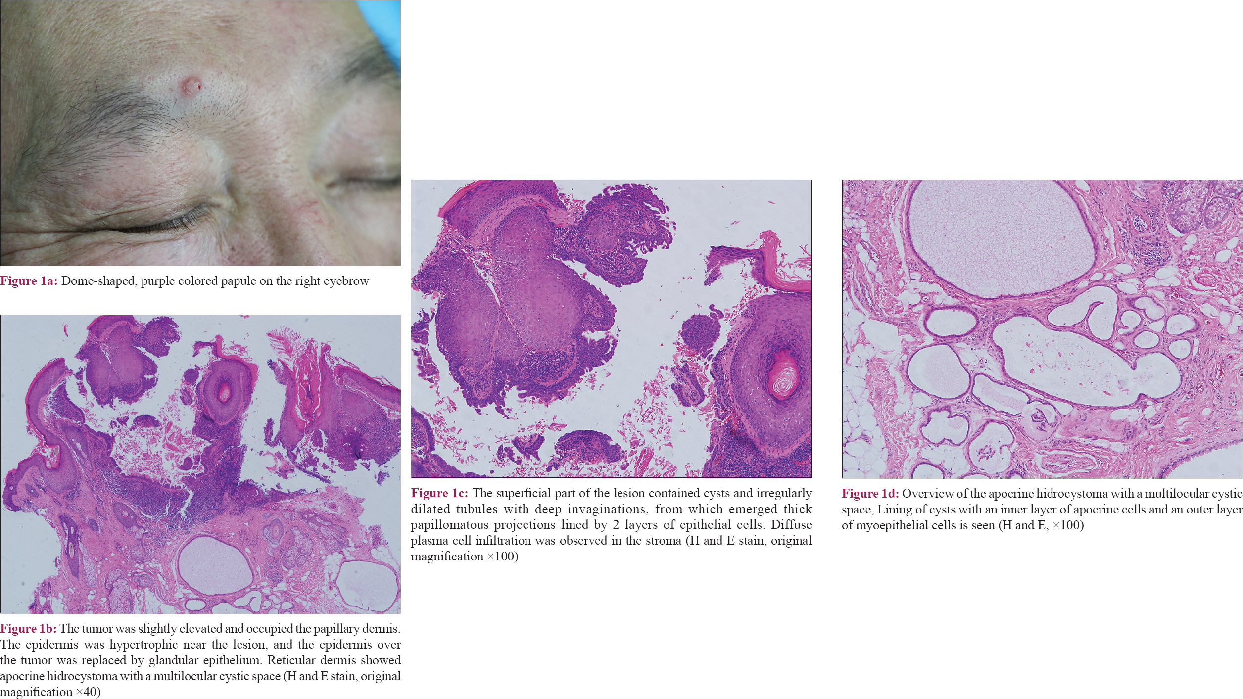
A 60-year-old Chinese male presented to our outpatient department with a solitary papular lesion on the right eyebrow. As stated by the patient, the lesion appeared 10 years back, as an asymptomatic, small, rounded lesion of skin color. Slowly the lesion grew bigger in size, and he felt an itch 4 days back. No other associated symptoms were reported. His medical history and family history were noncontributory. On physical examination, a purple-colored firm papule measuring approximately 5 mm × 5 mm with slight scaling on the surface was noted [Figure - 1]a. The surrounding skin appeared normal. There was no regional lymphadenopathy and no other skin lesions were noted elsewhere. Systemic examination revealed no abnormality. Complete excision was performed and the specimen was sent for histopathology examination, which showed that the superficial part of the lesion contained cysts and irregularly dilated tubules with deep invaginations, from which emerged thick papillomatous projections lined by two layers of epithelial cells. Diffuse plasma cell infiltration was observed in the stroma. The tumor was slightly elevated and occupied the papillary dermis. The epidermis was hypertrophic near the lesion, and the epidermis over the tumor was replaced by glandular epithelium. Reticular dermis showed apocrine hidrocystoma with a multilocular cystic space. Lining of cysts with an inner layer of apocrine cells and an outer layer of myoepithelial cells were noted [Figure - 1]b, [Figure - 1]c, [Figure - 1]d. Hence, a confirmatory diagnosis of syringocystadenoma papilliferum with coexisting hidrocystoma was made.
 |
| Figure 1: |
Syringocystadenoma occurs most commonly on the head and neck region.[1] Occurrence at unusual locations has been reported such as chest, abdomen, thigh, buttock, vulva, scrotum, pinna, eyelid, and external auditory canal.[2] It is usually associated with nevus sebaceus of Jadassohn but can arise de novo, as in our case. Fifty percent of these lesions are present at birth or in early childhood and 15–30% develop during puberty.[3] Of note, our patient developed this lesion at the age of 50 years. Moreover, since Pinkus first reported this benign neoplasm in 1954, its coexistence with other benign neoplasms has been reported by only a handful of studies.[4],[5],[6] We report this case because of its rarity.
Acknowledgment
This study was funded by grants from National Natural Science Foundation of China (NSFC 81360236), Shanghai hospital development center project (SHDC12014217) and Science and Technology commission of Shanghai municipality (16411961500).
Declaration of patient consent
The authors certify that they have obtained all appropriate patient consent forms. In the form, the patient has given his consent for his images and other clinical information to be reported in the journal. The patient understands that name and initials will not be published and due efforts will be made to conceal identity, but anonymity cannot be guaranteed.
Financial support and sponsorship
Nil.
Conflicts of interest
There are no conflicts of interest.
| 1. |
Muthusamy RK, Mehta SS. Syringocystadenocarcinoma papilliferum with coexisting trichoblastoma: A case report with review of literature. Indian J Dermatol Venereol Leprol 2017;83:574-6.
[Google Scholar]
|
| 2. |
Xu D, Bi T, Lan H, Yu W, Wang W, Cao F, et al. Syringocystadenoma papilliferum in the right lower abdomen: A case report and review of literature. Onco Targets Ther 2013;6:233-6.
[Google Scholar]
|
| 3. |
Böni R, Xin H, Hohl D, Panizzon R, Burg G. Syringocystadenoma papilliferum: A study of potential tumor suppressor genes. Am J Dermatopathol 2001;23:87-9.
[Google Scholar]
|
| 4. |
Karg E, Korom I, Varga E, Ban G, Turi S. Congenital syringocystadenoma papilliferum. Pediatr Dermatol 2008;25:132-3.
[Google Scholar]
|
| 5. |
Schewach-Millet M, Trau H. Congenital papillated apocrine cystadenoma: A mixed form of hidrocystoma, hidradenoma papilliferum, and syringocystadenoma papilliferum. J Am Acad Dermatol 1984;11:374-6.
[Google Scholar]
|
| 6. |
Arias-Santiago S, Aceituno-Madera P, Aneiros-Fernández J, Gutiérrez-Salmerón MT, Naranjo-Sintes R. Syringocystoadenoma papilliferum associated with apocrine hidrocystoma and verruca. Dermatol Online J 2009;15:9.
[Google Scholar]
|
Fulltext Views
2,678
PDF downloads
2,010


